当前位置:
X-MOL 学术
›
Acta Crystallogr. A Found. Adv.
›
论文详情
Our official English website, www.x-mol.net, welcomes your
feedback! (Note: you will need to create a separate account there.)
Electron image contrast analysis of mosaicity in rutile nanocrystals using direct electron detection
Acta Crystallographica Section A: Foundations and Advances ( IF 1.9 ) Pub Date : 2020-10-23 , DOI: 10.1107/s2053273320011055 Aram Yoon , Yu-Tsun Shao , Jane Howe , Jian-Min Zuo
Acta Crystallographica Section A: Foundations and Advances ( IF 1.9 ) Pub Date : 2020-10-23 , DOI: 10.1107/s2053273320011055 Aram Yoon , Yu-Tsun Shao , Jane Howe , Jian-Min Zuo
|
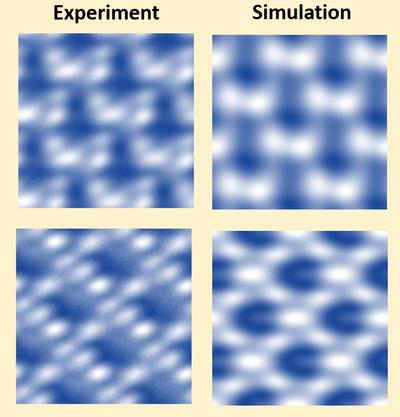
Direct electron detection provides high detective quantum efficiency, significantly improved point spread function and fast read‐out which have revolutionized the field of cryogenic electron microscopy. However, these benefits for high‐resolution electron microscopy (HREM) are much less exploited, especially for in situ study where major impacts on crystallographic structural studies could be made. By using direct detection in electron counting mode, rutile nanocrystals have been imaged at high temperature inside an environmental transmission electron microscope. The improvements in image contrast are quantified by comparison with a charge‐coupled device (CCD) camera and by image matching with simulations using an automated approach based on template matching. Together, these approaches enable a direct measurement of 3D shape and mosaicity (∼1°) of a vacuum‐reduced TiO2 nanocrystal about 50 nm in size. Thus, this work demonstrates the possibility of quantitative HREM image analysis based on direct electron detection.
中文翻译:

使用直接电子检测技术对金红石型纳米晶体中镶嵌性的电子图像对比度分析
直接电子检测提供了高检测量子效率,显着改善的点扩散功能和快速读取功能,这彻底改变了低温电子显微镜领域。但是,高分辨率电子显微术(HREM)的这些好处很少得到利用,尤其是在原位可以对晶体结构研究产生重大影响的研究。通过在电子计数模式下使用直接检测,金红石型纳米晶体已经在高温环境透射电子显微镜中成像。通过与电荷耦合器件(CCD)相机进行比较,并通过基于模板匹配的自动方法对图像进行仿真匹配,可以量化图像对比度的提高。总之,这些方法可以直接测量尺寸约50 nm的真空还原TiO 2纳米晶体的3D形状和镶嵌度(约1°)。因此,这项工作证明了基于直接电子检测的定量HREM图像分析的可能性。
更新日期:2020-11-02
中文翻译:
使用直接电子检测技术对金红石型纳米晶体中镶嵌性的电子图像对比度分析
直接电子检测提供了高检测量子效率,显着改善的点扩散功能和快速读取功能,这彻底改变了低温电子显微镜领域。但是,高分辨率电子显微术(HREM)的这些好处很少得到利用,尤其是在原位可以对晶体结构研究产生重大影响的研究。通过在电子计数模式下使用直接检测,金红石型纳米晶体已经在高温环境透射电子显微镜中成像。通过与电荷耦合器件(CCD)相机进行比较,并通过基于模板匹配的自动方法对图像进行仿真匹配,可以量化图像对比度的提高。总之,这些方法可以直接测量尺寸约50 nm的真空还原TiO 2纳米晶体的3D形状和镶嵌度(约1°)。因此,这项工作证明了基于直接电子检测的定量HREM图像分析的可能性。


 京公网安备 11010802027423号