当前位置:
X-MOL 学术
›
Microsc. Res. Tech.
›
论文详情
Our official English website, www.x-mol.net, welcomes your
feedback! (Note: you will need to create a separate account there.)
Suitability of the thawed algae for transmission electron microscopy study: Ultrastructural investigation on Coccomyxa melkonianii SCCA 048
Microscopy Research and Technique ( IF 2.0 ) Pub Date : 2020-10-23 , DOI: 10.1002/jemt.23626 Michela Isola 1 , Santina Soru 2 , Francesco Loy 1 , Veronica Malavasi 2 , Raffaella Isola 1 , Giacomo Cao 2, 3
Microscopy Research and Technique ( IF 2.0 ) Pub Date : 2020-10-23 , DOI: 10.1002/jemt.23626 Michela Isola 1 , Santina Soru 2 , Francesco Loy 1 , Veronica Malavasi 2 , Raffaella Isola 1 , Giacomo Cao 2, 3
Affiliation
|
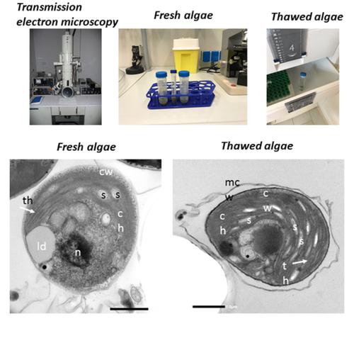
Morphological and ultrastructural investigations are crucial for the identification and characterization of species such as microalgae, microorganisms that greatly change their morphology and physiology during their life cycle. Transmission electron microscopy (TEM) is an excellent tool for the ultrastructural observation of cells and their components. To date, limited ultrastructural studies have been carried out on microalgae, due to the difficulties in sample preparation. The aim of this work is to establish an appropriate fixation method that allows to better preserve the algal ultrastructure and test the suitability of the thawed algae for TEM observation. Fresh and thawed algae (Coccomyxa melkonianii SCCA 048) were fixed with different TEM fixation methods (a mix of glutaraldehyde and paraformaldehyde for several incubation times, sometimes preceded by a prefixation in cold methanol). The ultrastructural images obtained from fresh algae were compared to those obtained from frozen biomass. The best morphological results were achieved by fixing fresh algae in 1% paraformaldehyde and 1.25% glutaraldehyde for 5 hr. Pretreating with frozen methyl alcohol reduced fixation time to 2 hr. Both fresh and frozen algae ultrastructure were rather well preserved also with 1% paraformaldehyde and 1.25% glutaraldehyde for 2 hr. Ultrastuctural morphological images of the thawed algae demonstrated that also frozen samples can be used in TEM research, widening specimen suitability by means of this technique.
中文翻译:

解冻藻类在透射电子显微镜研究中的适用性:Coccomyxa melkonianii SCCA 048 的超微结构研究
形态学和超微结构研究对于微藻、微生物等物种的鉴定和表征至关重要,这些微生物在其生命周期中会极大地改变其形态和生理学。透射电子显微镜 (TEM) 是对细胞及其成分进行超微结构观察的绝佳工具。迄今为止,由于样品制备困难,已对微藻进行了有限的超微结构研究。这项工作的目的是建立一种适当的固定方法, 可以更好地保存藻类超微结构, 并测试解冻藻类对 TEM 观察的适用性。新鲜和解冻的藻类 ( Coccomyxa melkonianiiSCCA 048) 用不同的 TEM 固定方法固定(戊二醛和多聚甲醛的混合物多次孵育,有时在冷甲醇中加前缀)。将从新鲜藻类获得的超微结构图像与从冷冻生物质获得的超微结构图像进行比较。将新鲜藻类在 1% 多聚甲醛和 1.25% 戊二醛中固定 5 小时可获得最佳形态学结果。用冷冻甲醇预处理将固定时间缩短至 2 小时。新鲜的和冷冻的藻类超微结构也用 1% 多聚甲醛和 1.25% 戊二醛保存 2 小时。解冻藻类的超微结构形态图像表明,冷冻样品也可用于 TEM 研究,通过该技术扩大样品适用性。
更新日期:2020-10-23
中文翻译:
解冻藻类在透射电子显微镜研究中的适用性:Coccomyxa melkonianii SCCA 048 的超微结构研究
形态学和超微结构研究对于微藻、微生物等物种的鉴定和表征至关重要,这些微生物在其生命周期中会极大地改变其形态和生理学。透射电子显微镜 (TEM) 是对细胞及其成分进行超微结构观察的绝佳工具。迄今为止,由于样品制备困难,已对微藻进行了有限的超微结构研究。这项工作的目的是建立一种适当的固定方法, 可以更好地保存藻类超微结构, 并测试解冻藻类对 TEM 观察的适用性。新鲜和解冻的藻类 ( Coccomyxa melkonianiiSCCA 048) 用不同的 TEM 固定方法固定(戊二醛和多聚甲醛的混合物多次孵育,有时在冷甲醇中加前缀)。将从新鲜藻类获得的超微结构图像与从冷冻生物质获得的超微结构图像进行比较。将新鲜藻类在 1% 多聚甲醛和 1.25% 戊二醛中固定 5 小时可获得最佳形态学结果。用冷冻甲醇预处理将固定时间缩短至 2 小时。新鲜的和冷冻的藻类超微结构也用 1% 多聚甲醛和 1.25% 戊二醛保存 2 小时。解冻藻类的超微结构形态图像表明,冷冻样品也可用于 TEM 研究,通过该技术扩大样品适用性。


 京公网安备 11010802027423号