当前位置:
X-MOL 学术
›
J. Zool. Syst. Evol. Res.
›
论文详情
Our official English website, www.x-mol.net, welcomes your
feedback! (Note: you will need to create a separate account there.)
Ultrastructure of ganglia in the brachiopod Coptothyris grayi and its phylogenetic significance
Journal of Zoological Systematics and Evolutionary Research ( IF 2.0 ) Pub Date : 2020-10-21 , DOI: 10.1111/jzs.12427 Tatyana Kuzmina 1 , Elena Temereva 1, 2
Journal of Zoological Systematics and Evolutionary Research ( IF 2.0 ) Pub Date : 2020-10-21 , DOI: 10.1111/jzs.12427 Tatyana Kuzmina 1 , Elena Temereva 1, 2
Affiliation
|
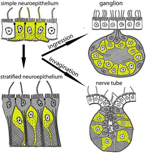
Since the study of the neuroarchitecture has phylogenetic value, it is important to describe the nervous system in poor investigated groups. We describe the ultrastructure of the supraenteric and subenteric ganglia in the rhynchonelliform brachiopod Coptothyris grayi. We found that the supraenteric ganglion is a neuroepithelium that consists of monociliated neurons among glial cells and a basal neuropil. The subenteric ganglion contains neurons of two morphologically distinct types and is represented by a stratified neuroepithelium with three layers. The apical layer is formed by the somata of glial cells. The middle layer contains the large perikarya of the neurons of the first type. The inner basal layer consists of a neuropil, basal projections of glial cells, and neurons of the second type. The brachiopod supraenteric ganglion exhibits the features of the simple neuroepithelium that is probably the plesiomorphic condition in the bilaterian ancestor. The brachiopod subenteric ganglion is a stratified neuroepithelium that has been described in some groups of protostomes and deuterostomes. The next step in the evolution of the invertebrate nervous system was an internalization. In many groups of protostomes, the subepidermal ganglia, which consist of perikarya surrounding a central neuropil, are formed by ingression of the neuroepithelium. In deuterostomes, the nerve tube with an inner neural canal is formed by invagination of the stratified neuroepithelium. Thus, the so‐called ganglia of brachiopods are neuroepithelia that have retained the plesiomorphic condition of the bilaterian nervous system and that cannot be regarded as true ganglia.
中文翻译:

腕足类Coptothyris grayi神经节的超微结构及其系统发育意义
由于对神经体系结构的研究具有系统发育价值,因此在贫穷的研究人群中描述神经系统很重要。我们描述了在上颌和下肠神经节的超细结构。我们发现肠上神经节是神经上皮,由神经胶质细胞和基底神经纤维之间的单纤毛神经元组成。肠下神经节包含两种形态上不同的神经元,并由具有三层的分层神经上皮表示。根尖层由神经胶质细胞的体细胞形成。中间层包含第一类神经元的大周核。内基底层由神经纤维,神经胶质细胞的基底突起和第二类型的神经元组成。腕足类上肠神经节表现出简单的神经上皮的特征,这可能是在双侧祖先的多形性状态。腕足动物肠下神经节是一种分层的神经上皮,已在某些组的原虫和氘核口中进行了描述。无脊椎动物神经系统进化的下一步是内在化。在许多原虫组中,表皮下神经节由神经上皮的侵入形成,其由围绕中央神经纤维的周围核构成。在氘化子宫口中,通过分层神经上皮的内陷形成具有神经内管的神经管。因此,所谓的腕足神经节是神经上皮细胞,保留了双侧神经系统的多形性状态,不能被视为真正的神经节。内层神经管的神经管是通过分层神经上皮的内陷形成的。因此,所谓的腕足神经节是神经上皮细胞,保留了双侧神经系统的多形性状态,不能被视为真正的神经节。内层神经管的神经管是通过分层神经上皮的内陷形成的。因此,所谓的腕足神经节是神经上皮细胞,保留了双侧神经系统的多形性状态,不能被视为真正的神经节。
更新日期:2020-10-21
中文翻译:
腕足类Coptothyris grayi神经节的超微结构及其系统发育意义
由于对神经体系结构的研究具有系统发育价值,因此在贫穷的研究人群中描述神经系统很重要。我们描述了在上颌和下肠神经节的超细结构。我们发现肠上神经节是神经上皮,由神经胶质细胞和基底神经纤维之间的单纤毛神经元组成。肠下神经节包含两种形态上不同的神经元,并由具有三层的分层神经上皮表示。根尖层由神经胶质细胞的体细胞形成。中间层包含第一类神经元的大周核。内基底层由神经纤维,神经胶质细胞的基底突起和第二类型的神经元组成。腕足类上肠神经节表现出简单的神经上皮的特征,这可能是在双侧祖先的多形性状态。腕足动物肠下神经节是一种分层的神经上皮,已在某些组的原虫和氘核口中进行了描述。无脊椎动物神经系统进化的下一步是内在化。在许多原虫组中,表皮下神经节由神经上皮的侵入形成,其由围绕中央神经纤维的周围核构成。在氘化子宫口中,通过分层神经上皮的内陷形成具有神经内管的神经管。因此,所谓的腕足神经节是神经上皮细胞,保留了双侧神经系统的多形性状态,不能被视为真正的神经节。内层神经管的神经管是通过分层神经上皮的内陷形成的。因此,所谓的腕足神经节是神经上皮细胞,保留了双侧神经系统的多形性状态,不能被视为真正的神经节。内层神经管的神经管是通过分层神经上皮的内陷形成的。因此,所谓的腕足神经节是神经上皮细胞,保留了双侧神经系统的多形性状态,不能被视为真正的神经节。


 京公网安备 11010802027423号