当前位置:
X-MOL 学术
›
ACS Chem. Biol.
›
论文详情
Our official English website, www.x-mol.net, welcomes your
feedback! (Note: you will need to create a separate account there.)
Visualizing HIF-1α mRNA in a Subpopulation of Bone Marrow-Derived Cells to Predict Retinal Neovascularization
ACS Chemical Biology ( IF 3.5 ) Pub Date : 2020-10-20 , DOI: 10.1021/acschembio.0c00662 Md. Imam Uddin 1, 2 , Tyler C. Kilburn 1 , Craig L. Duvall 2 , John S. Penn 1, 3, 4
ACS Chemical Biology ( IF 3.5 ) Pub Date : 2020-10-20 , DOI: 10.1021/acschembio.0c00662 Md. Imam Uddin 1, 2 , Tyler C. Kilburn 1 , Craig L. Duvall 2 , John S. Penn 1, 3, 4
Affiliation

|
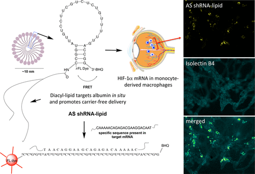
Bone marrow-derived progenitor cells and macrophages are known to migrate into the retina in response to inflammation and neovascularization. These migratory cells might play important regulatory roles in the pathogenesis of neovascularization, a common complication observed in diabetic retinopathy, retinopathy of prematurity, and retinal vein occlusion. Hypoxia-inducible factor 1α (HIF-1α) has been shown to contribute to the pathogenesis of retinal inflammation and neovascularization. However, contributions of monocyte-derived macrophages to neovascularization are largely unknown. We hypothesized that selective visualization of these microglia/macrophages could be a powerful method for predicting the onset of neovascularization and its progression at the molecular level. In this report, we describe the synthesis of a new hybrid nanoparticle to visualize HIF-1α mRNA selectively in microglia/macrophages in a mouse model of neovascularization. HIF-1α expression was confirmed in MRC-1 positive monocytes/macrophages as well as in CD4 positive T-cells and CD19 positive B-cells using single-cell RNA sequencing data analysis. The imaging probes (AS- or NS-shRNA-lipid) were synthesized by conjugating diacyl-lipids to short hairpin RNA with an antisense sequence complementary to HIF-1α mRNA and a fluorophore that is quenched by a black hole quencher. We believe that imaging mRNA selectively in tissue specific microglia/macrophages could be a powerful method for predicting the onset of neovascularization, its progression, and its response to therapy.
中文翻译:

可视化骨髓衍生细胞亚群中的HIF-1αmRNA,以预测视网膜新血管形成
已知骨髓来源的祖细胞和巨噬细胞响应炎症和新血管形成而迁移到视网膜中。这些迁移细胞可能在新生血管形成的发病机理中起重要的调节作用,新生血管形成是糖尿病性视网膜病变,早产儿视网膜病变和视网膜静脉阻塞中常见的并发症。缺氧诱导因子1α(HIF-1α)已被证明与视网膜炎症和新生血管形成有关。然而,单核细胞衍生的巨噬细胞对新血管形成的贡献在很大程度上是未知的。我们假设这些小胶质细胞/巨噬细胞的选择性可视化可能是预测新血管形成及其分子水平进展的有效方法。在这份报告中使用单细胞RNA测序数据分析,在MRC-1阳性单核细胞/巨噬细胞以及CD4阳性T细胞和CD19阳性B细胞中证实了HIF-1α表达。成像探针(AS-或NS-shRNA-脂质)是通过将二酰基脂质与具有与HIF-1αmRNA互补的反义序列和被黑洞淬灭剂淬灭的荧光团的短发夹RNA缀合而合成的。我们认为,在组织特异性小胶质细胞/巨噬细胞中选择性地对mRNA成像可能是预测新血管形成,其进展及其对治疗反应的有力方法。
更新日期:2020-11-21
中文翻译:

可视化骨髓衍生细胞亚群中的HIF-1αmRNA,以预测视网膜新血管形成
已知骨髓来源的祖细胞和巨噬细胞响应炎症和新血管形成而迁移到视网膜中。这些迁移细胞可能在新生血管形成的发病机理中起重要的调节作用,新生血管形成是糖尿病性视网膜病变,早产儿视网膜病变和视网膜静脉阻塞中常见的并发症。缺氧诱导因子1α(HIF-1α)已被证明与视网膜炎症和新生血管形成有关。然而,单核细胞衍生的巨噬细胞对新血管形成的贡献在很大程度上是未知的。我们假设这些小胶质细胞/巨噬细胞的选择性可视化可能是预测新血管形成及其分子水平进展的有效方法。在这份报告中使用单细胞RNA测序数据分析,在MRC-1阳性单核细胞/巨噬细胞以及CD4阳性T细胞和CD19阳性B细胞中证实了HIF-1α表达。成像探针(AS-或NS-shRNA-脂质)是通过将二酰基脂质与具有与HIF-1αmRNA互补的反义序列和被黑洞淬灭剂淬灭的荧光团的短发夹RNA缀合而合成的。我们认为,在组织特异性小胶质细胞/巨噬细胞中选择性地对mRNA成像可能是预测新血管形成,其进展及其对治疗反应的有力方法。


 京公网安备 11010802027423号