Our official English website, www.x-mol.net, welcomes your
feedback! (Note: you will need to create a separate account there.)
Protein kinase C eta is activated in reactive astrocytes of an Alzheimer's disease mouse model: Evidence for its immunoregulatory function in primary astrocytes
Glia ( IF 5.4 ) Pub Date : 2020-10-17 , DOI: 10.1002/glia.23921 Amitha Muraleedharan 1, 2 , Noa Rotem-Dai 1 , Itai Strominger 1, 2 , Nikhil Ponnoor Anto 1 , Noah Isakov 1 , Alon Monsonego 1, 2 , Etta Livneh 1
Glia ( IF 5.4 ) Pub Date : 2020-10-17 , DOI: 10.1002/glia.23921 Amitha Muraleedharan 1, 2 , Noa Rotem-Dai 1 , Itai Strominger 1, 2 , Nikhil Ponnoor Anto 1 , Noah Isakov 1 , Alon Monsonego 1, 2 , Etta Livneh 1
Affiliation
|
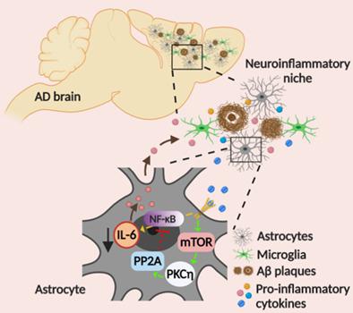
Alzheimer's disease (AD) is the primary cause of age‐related dementia. Pathologically, AD is characterized by synaptic loss, the accumulation of β‐amyloid peptides and neurofibrillary tangles, glial activation, and neuroinflammation. Whereas extensive studies focused on neurons and activation of microglia in AD, the role of astrocytes has not been well‐characterized. Protein kinase C (PKC) was also implicated in AD; however, its role in astrocyte activation was not elucidated. Using the 5XFAD mouse model of AD, we show that PKC‐eta (PKCη), an astrocyte‐specific stress‐activated and anti‐apoptotic kinase, plays a role in reactive astrocytes. We demonstrate that PKCη staining is highly enriched in cortical astrocytes in a disease‐dependent manner and in the vicinity of amyloid‐β peptides plaques. Moreover, activation of PKCη, as indicated by its increased phosphorylation levels, is exhibited mainly in cortical astrocytes derived from adult 5XFAD mice. PKCη activation was associated with elevated levels of reactive astrocytic markers and upregulation of the pro‐inflammatory cytokine interleukin 6 (IL‐6) compared to littermate controls. Notably, inhibiting the kinase activity of PKCη in 5XFAD astrocyte cultures markedly increased the levels of secreted IL‐6—a phenomenon that was also observed in wild‐type astrocytes stimulated by inflammatory cytokines (e.g., TNFα, IL‐1). Similar increase in the release of IL‐6 was also observed upon inhibition of either the mammalian target of rapamycin (mTOR) or the protein phosphatase 2A (PP2A). Our findings suggest that the mTOR‐PKCη‐PP2A signaling cascade functions as a negative feedback loop of NF‐κB‐induced IL‐6 release in astrocytes. Thus, we identify PKCη as a regulator of neuroinflammation in AD.
中文翻译:

蛋白激酶 C eta 在阿尔茨海默病小鼠模型的反应性星形胶质细胞中被激活:其在原代星形胶质细胞中的免疫调节功能的证据
阿尔茨海默病 (AD) 是导致年龄相关性痴呆的主要原因。在病理学上,AD 的特征是突触缺失、β-淀粉样肽和神经原纤维缠结的积累、神经胶质激活和神经炎症。尽管广泛的研究集中在 AD 中的神经元和小胶质细胞的激活,但星形胶质细胞的作用尚未得到很好的表征。蛋白激酶 C (PKC) 也与 AD 相关。然而,它在星形胶质细胞活化中的作用尚未阐明。使用 AD 的 5XFAD 小鼠模型,我们发现 PKC-eta (PKCη),一种星形胶质细胞特异性应激激活和抗凋亡激酶,在反应性星形胶质细胞中发挥作用。我们证明了 PKCη 染色在皮质星形胶质细胞中以疾病依赖性方式和淀粉样蛋白-β 肽斑块附近高度富集。此外,激活 PKCη,如其增加的磷酸化水平所示,主要表现在源自成年 5XFAD 小鼠的皮质星形胶质细胞中。与同窝对照组相比,PKCη 激活与反应性星形胶质细胞标志物水平升高和促炎细胞因子白细胞介素 6 (IL-6) 上调有关。值得注意的是,在 5XFAD 星形胶质细胞培养物中抑制 PKCη 的激酶活性显着增加了分泌的 IL-6 水平——这种现象在受炎性细胞因子(如 TNFα、IL-1)刺激的野生型星形胶质细胞中也观察到。在抑制哺乳动物雷帕霉素靶标 (mTOR) 或蛋白磷酸酶 2A (PP2A) 后,也观察到 IL-6 释放的类似增加。我们的研究结果表明,mTOR-PKCη-PP2A 信号级联作为 NF-κB 诱导的星形胶质细胞释放 IL-6 的负反馈回路发挥作用。因此,我们将 PKCη 确定为 AD 中神经炎症的调节剂。
更新日期:2020-10-17
中文翻译:
蛋白激酶 C eta 在阿尔茨海默病小鼠模型的反应性星形胶质细胞中被激活:其在原代星形胶质细胞中的免疫调节功能的证据
阿尔茨海默病 (AD) 是导致年龄相关性痴呆的主要原因。在病理学上,AD 的特征是突触缺失、β-淀粉样肽和神经原纤维缠结的积累、神经胶质激活和神经炎症。尽管广泛的研究集中在 AD 中的神经元和小胶质细胞的激活,但星形胶质细胞的作用尚未得到很好的表征。蛋白激酶 C (PKC) 也与 AD 相关。然而,它在星形胶质细胞活化中的作用尚未阐明。使用 AD 的 5XFAD 小鼠模型,我们发现 PKC-eta (PKCη),一种星形胶质细胞特异性应激激活和抗凋亡激酶,在反应性星形胶质细胞中发挥作用。我们证明了 PKCη 染色在皮质星形胶质细胞中以疾病依赖性方式和淀粉样蛋白-β 肽斑块附近高度富集。此外,激活 PKCη,如其增加的磷酸化水平所示,主要表现在源自成年 5XFAD 小鼠的皮质星形胶质细胞中。与同窝对照组相比,PKCη 激活与反应性星形胶质细胞标志物水平升高和促炎细胞因子白细胞介素 6 (IL-6) 上调有关。值得注意的是,在 5XFAD 星形胶质细胞培养物中抑制 PKCη 的激酶活性显着增加了分泌的 IL-6 水平——这种现象在受炎性细胞因子(如 TNFα、IL-1)刺激的野生型星形胶质细胞中也观察到。在抑制哺乳动物雷帕霉素靶标 (mTOR) 或蛋白磷酸酶 2A (PP2A) 后,也观察到 IL-6 释放的类似增加。我们的研究结果表明,mTOR-PKCη-PP2A 信号级联作为 NF-κB 诱导的星形胶质细胞释放 IL-6 的负反馈回路发挥作用。因此,我们将 PKCη 确定为 AD 中神经炎症的调节剂。


 京公网安备 11010802027423号