当前位置:
X-MOL 学术
›
Brain Pathol.
›
论文详情
Our official English website, www.x-mol.net, welcomes your
feedback! (Note: you will need to create a separate account there.)
Overexpression of the ubiquitin‐editing enzyme A20 in the brain lesions of Multiple Sclerosis patients: moving from systemic to central nervous system inflammation
Brain Pathology ( IF 5.8 ) Pub Date : 2020-10-14 , DOI: 10.1111/bpa.12906 Simona Perga 1, 2, 3 , Francesca Montarolo 1, 2, 4 , Serena Martire 1, 2 , Brigitta Bonaldo 1, 3 , Gabriele Bono 1, 2 , Jessica Bertolo 1, 2 , Roberta Magliozzi 5, 6 , Antonio Bertolotto 1, 2
Brain Pathology ( IF 5.8 ) Pub Date : 2020-10-14 , DOI: 10.1111/bpa.12906 Simona Perga 1, 2, 3 , Francesca Montarolo 1, 2, 4 , Serena Martire 1, 2 , Brigitta Bonaldo 1, 3 , Gabriele Bono 1, 2 , Jessica Bertolo 1, 2 , Roberta Magliozzi 5, 6 , Antonio Bertolotto 1, 2
Affiliation
|
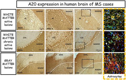
Multiple Sclerosis (MS) is a chronic demyelinating disease of the central nervous system (CNS) in which inflammation plays a key pathological role. Recent evidences showed that systemic inflammation induces increasing cell infiltration within meninges and perivascular spaces in the brain parenchyma, triggering resident microglial and astrocytic activation. The anti‐inflammatory enzyme A20, also named TNF associated protein 3 (TNFAIP3), is considered a central gatekeeper in inflammation and peripheral immune system regulation through the inhibition of NF‐kB. The TNFAIP3 locus is genetically associated to MS and its transcripts is downregulated in blood cells in treatment‐naïve MS patients. Recently, several evidences in mouse models have led to hypothesize a function of A20 also in the CNS. Thus, here we aimed to unveil a possible contribution of A20 to the CNS human MS pathology. By immunohistochemistry/immunofluorescence and biomolecular techniques on post‐mortem brain tissue blocks obtained from control cases (CC) and progressive MS cases, we demonstrated that A20 is present in CC brain tissues in both white matter (WM) regions, mainly in few parenchymal astrocytes, and in grey matter (GM) areas, in some neuronal populations. Conversely, in MS brain tissues, we observed increased expression of A20 by perivascular infiltrating macrophages, resident‐activated astrocytes, and microglia in all the active and chronic active WM lesions. A20 was highly expressed also in the majority of active cortical lesions compared to the neighboring areas of normal‐appearing grey matter (NAGM) and control GM, particularly by activated astrocytes. We demonstrated increased A20 expression in the active MS plaques, particularly in macrophages and resident astrocytes, suggesting a key role of this molecule in chronic inflammation.
中文翻译:

泛素编辑酶 A20 在多发性硬化症患者脑损伤中的过度表达:从全身性炎症到中枢神经系统炎症
多发性硬化症 (MS) 是一种慢性中枢神经系统 (CNS) 脱髓鞘疾病,其中炎症起着关键的病理作用。最近的证据表明,全身性炎症会导致脑实质内脑膜和血管周围间隙的细胞浸润增加,从而引发常驻小胶质细胞和星形胶质细胞的激活。抗炎酶 A20,也称为 TNF 相关蛋白 3(TNFAIP3),被认为是通过抑制 NF-kB 来调节炎症和外周免疫系统的中心守门人。TNFAIP3 基因座与 MS 遗传相关,其转录本在未接受治疗的 MS 患者的血细胞中下调。最近,小鼠模型中的一些证据导致假设 A20 在 CNS 中也有功能。因此,在这里,我们旨在揭示 A20 对 CNS 人类 MS 病理学的可能贡献。通过免疫组织化学/免疫荧光和生物分子技术验尸从对照病例 (CC) 和进展性 MS 病例获得的脑组织块,我们证明 A20 存在于 CC 脑组织的两个白质 (WM) 区域,主要存在于少数实质星形胶质细胞和灰质 (GM) 区域,在一些神经元群。相反,在 MS 脑组织中,我们观察到血管周围浸润的巨噬细胞、驻留激活的星形胶质细胞和所有活动性和慢性活动性 WM 病变中的小胶质细胞的 A20 表达增加。与外观正常的灰质 (NAGM) 和对照 GM 的邻近区域相比,A20 在大多数活动性皮层病变中也高度表达,尤其是在活化的星形胶质细胞中。我们证明活性 MS 斑块中 A20 表达增加,特别是在巨噬细胞和常驻星形胶质细胞中,
更新日期:2020-10-14
中文翻译:
泛素编辑酶 A20 在多发性硬化症患者脑损伤中的过度表达:从全身性炎症到中枢神经系统炎症
多发性硬化症 (MS) 是一种慢性中枢神经系统 (CNS) 脱髓鞘疾病,其中炎症起着关键的病理作用。最近的证据表明,全身性炎症会导致脑实质内脑膜和血管周围间隙的细胞浸润增加,从而引发常驻小胶质细胞和星形胶质细胞的激活。抗炎酶 A20,也称为 TNF 相关蛋白 3(TNFAIP3),被认为是通过抑制 NF-kB 来调节炎症和外周免疫系统的中心守门人。TNFAIP3 基因座与 MS 遗传相关,其转录本在未接受治疗的 MS 患者的血细胞中下调。最近,小鼠模型中的一些证据导致假设 A20 在 CNS 中也有功能。因此,在这里,我们旨在揭示 A20 对 CNS 人类 MS 病理学的可能贡献。通过免疫组织化学/免疫荧光和生物分子技术验尸从对照病例 (CC) 和进展性 MS 病例获得的脑组织块,我们证明 A20 存在于 CC 脑组织的两个白质 (WM) 区域,主要存在于少数实质星形胶质细胞和灰质 (GM) 区域,在一些神经元群。相反,在 MS 脑组织中,我们观察到血管周围浸润的巨噬细胞、驻留激活的星形胶质细胞和所有活动性和慢性活动性 WM 病变中的小胶质细胞的 A20 表达增加。与外观正常的灰质 (NAGM) 和对照 GM 的邻近区域相比,A20 在大多数活动性皮层病变中也高度表达,尤其是在活化的星形胶质细胞中。我们证明活性 MS 斑块中 A20 表达增加,特别是在巨噬细胞和常驻星形胶质细胞中,


 京公网安备 11010802027423号