Journal of Neuroradiology ( IF 3.0 ) Pub Date : 2020-10-13 , DOI: 10.1016/j.neurad.2020.09.008 Hakan Cebeci 1 , Abidin Kilincer 1 , Halil İbrahim Duran 1 , Nusret Seher 1 , Mert Şahinoğlu 2 , Hakan Karabağlı 2 , Pınar Karabağlı 3 , Yahya Paksoy 4
|
Background and purpose
Meningiomas and schwannomas are common extra-axial brain tumors. Discrimination is challenging in some locations when characteristic imaging features are absent. This study investigated the accuracy of percentage signal recoveries obtained from dynamic susceptibility contrast perfusion imaging (DSC-PI) in discriminating meningiomas and schwannomas.
Material and methods
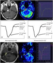
Retrospective database research was conducted. Sixty nine meningioma and 15 schwannoma having DSC-PI between January 2016 and February 2020 were included. Time to signal intensity curves (TSIC) were analyzed and grouped as T1-dominant leakage, T2*-dominant leakage and return to baseline. Relative cerebral blood volume (rCBV), relative mean transit time (rMTT), percentage signal recovery 1 (PSR 1) and PSR 2 values were calculated. The differences between the groups were investigated. Receiver operating characteristic curves were operated.
Results
rCBV, rMTT, PSR 1 and PSR 2 values were statistically different between meningiomas and schwannomas. PSR 2 provided the best discrimination. With the cut off value of 1.08 for PSR 2, meningiomas and schwannomas were differentiated with 95.7% sensitivity and 93.3% specificity. TSICs were also different between two groups. Most of meningiomas showed T2*-dominant leakage (78.2%), whereas most of shwannomas showed T1-dominant leakage (93.3%).
Conclusion
DSC-PI is a useful imaging tool for non-invasive discrimination of meningiomas and schwannomas. Particularly, percentage signal recoveries discriminates meningiomas and schwannomas with high sensitivity and specificity.
中文翻译:
使用时间-信号强度曲线和动态磁敏灌注成像获得的信号恢复百分比精确区分脑膜瘤和神经鞘瘤
背景和目的
脑膜瘤和神经鞘瘤是常见的轴外脑肿瘤。当特征成像特征不存在时,在某些位置区分是具有挑战性的。本研究调查了从动态磁敏感对比灌注成像 (DSC-PI) 获得的信号恢复百分比在区分脑膜瘤和神经鞘瘤方面的准确性。
材料与方法
进行了回顾性数据库研究。包括 2016 年 1 月至 2020 年 2 月期间患有 DSC-PI 的 69 例脑膜瘤和 15 例神经鞘瘤。分析信号时间强度曲线 (TSIC) 并将其分组为 T1 显性渗漏、T2* 显性渗漏和返回基线。计算相对脑血容量 (rCBV)、相对平均传输时间 (rMTT)、信号恢复百分比 1 (PSR 1) 和 PSR 2 值。研究了组之间的差异。操作接受者操作特征曲线。
结果
rCBV、rMTT、PSR 1 和 PSR 2 值在脑膜瘤和神经鞘瘤之间具有统计学差异。PSR 2 提供了最好的辨别力。PSR 2 的截止值为 1.08,区分脑膜瘤和神经鞘瘤的敏感性为 95.7%,特异性为 93.3%。两组之间的 TSIC 也不同。大多数脑膜瘤显示 T2* 主导的渗漏 (78.2%),而大多数神经鞘瘤显示 T1 主导的渗漏 (93.3%)。
结论
DSC-PI 是一种有用的成像工具,可用于非侵入性区分脑膜瘤和神经鞘瘤。特别是,百分比信号恢复以高灵敏度和特异性区分脑膜瘤和神经鞘瘤。


 京公网安备 11010802027423号