当前位置:
X-MOL 学术
›
Microsc. Res. Tech.
›
论文详情
Our official English website, www.x-mol.net, welcomes your
feedback! (Note: you will need to create a separate account there.)
Application of Scanning Electron Microscopy in the observation of dentin‐adhesive interface
Microscopy Research and Technique ( IF 2.0 ) Pub Date : 2020-10-12 , DOI: 10.1002/jemt.23618 Bojana D Ramić 1 , Igor Lj Stojanac 1 , Milan R Drobac 1 , Ivana R Kantardžić 1 , Aleksandra Z Maletin 1 , Milica T Cvjetićanin 1 , Katarina S Otašević 1 , Ljubomir M Petrović 1
Microscopy Research and Technique ( IF 2.0 ) Pub Date : 2020-10-12 , DOI: 10.1002/jemt.23618 Bojana D Ramić 1 , Igor Lj Stojanac 1 , Milan R Drobac 1 , Ivana R Kantardžić 1 , Aleksandra Z Maletin 1 , Milica T Cvjetićanin 1 , Katarina S Otašević 1 , Ljubomir M Petrović 1
Affiliation
|
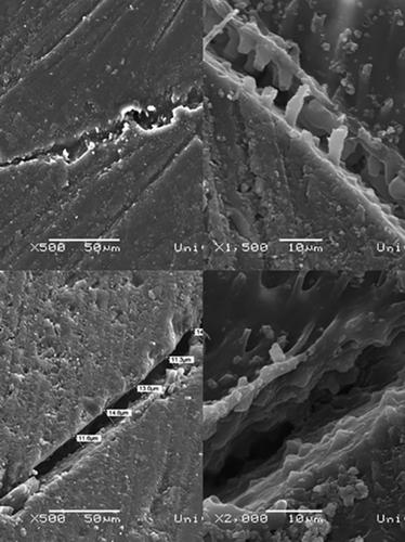
The objective of this study was observation of the adhesive interface on original tooth samples, as well as their epoxy replicas, under SEM. A light‐cure flowable composite was incrementally placed and light‐polymerized in previously prepared cylindrical dentinal cavities on the buccal surfaces of extracted human third molars. After finishing procedures, impressions of the composite/dentin margin were made using polyvinylsiloxane in order to obtain accurate epoxy replicas for SEM analysis. Ultrastructural morphology of the adhesive surface was observed at high magnifications (≥1,000×) on original tooth samples, which were previously prepared to expose the part of the dentin surface, which participates in the formation of adhesive bond. SEM micrographs showed that marginal adaptation was mostly of acceptable quality. In some of the SEM micrographs of original tooth samples, marginal gap formation, and resin tag breakdown were noted, which were ascribed to polymerization shrinkage. Profound understanding of ultrastructural morphology is necessary for achieving more predictable and durable margin between composite restorations and surrounding tooth structures, and SEM analysis can serve that purpose.
中文翻译:

扫描电镜在牙本质-胶粘剂界面观察中的应用
本研究的目的是在 SEM 下观察原始牙齿样品及其环氧树脂复制品上的粘合界面。将光固化可流动复合材料逐渐放置并光聚合在先前制备的圆柱形牙本质腔中,该腔位于拔出的人类第三磨牙的颊面。完成程序后,使用聚乙烯硅氧烷制作复合材料/牙本质边缘的印模,以获得用于 SEM 分析的准确环氧树脂复制品。在原始牙齿样品上以高放大倍数(≥1,000 倍)观察粘合剂表面的超微结构形态,这些样品预先制备以暴露参与粘合剂形成的牙本质表面部分。SEM 显微照片显示边缘适应大多具有可接受的质量。在原始牙齿样品的一些 SEM 显微照片中,注意到边缘间隙形成和树脂标签破裂,这归因于聚合收缩。深入了解超微结构形态对于在复合修复体和周围牙齿结构之间实现更可预测和更持久的边缘是必要的,而 SEM 分析可以达到这一目的。
更新日期:2020-10-12
中文翻译:
扫描电镜在牙本质-胶粘剂界面观察中的应用
本研究的目的是在 SEM 下观察原始牙齿样品及其环氧树脂复制品上的粘合界面。将光固化可流动复合材料逐渐放置并光聚合在先前制备的圆柱形牙本质腔中,该腔位于拔出的人类第三磨牙的颊面。完成程序后,使用聚乙烯硅氧烷制作复合材料/牙本质边缘的印模,以获得用于 SEM 分析的准确环氧树脂复制品。在原始牙齿样品上以高放大倍数(≥1,000 倍)观察粘合剂表面的超微结构形态,这些样品预先制备以暴露参与粘合剂形成的牙本质表面部分。SEM 显微照片显示边缘适应大多具有可接受的质量。在原始牙齿样品的一些 SEM 显微照片中,注意到边缘间隙形成和树脂标签破裂,这归因于聚合收缩。深入了解超微结构形态对于在复合修复体和周围牙齿结构之间实现更可预测和更持久的边缘是必要的,而 SEM 分析可以达到这一目的。


 京公网安备 11010802027423号