当前位置:
X-MOL 学术
›
J. Biophotonics
›
论文详情
Our official English website, www.x-mol.net, welcomes your feedback! (Note: you will need to create a separate account there.)
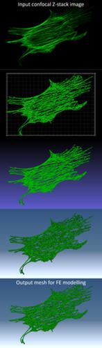
3D immuno‐confocal image reconstruction of fibroblast cytoskeleton and nucleus architecture
Journal of Biophotonics ( IF 2.8 ) Pub Date : 2020-10-10 , DOI: 10.1002/jbio.202000202 Petar Markov 1, 2, 3 , Anthony J Hayes 4 , Hanxing Zhu 3 , Craig Boote 2 , Emma J Blain 1, 5
Journal of Biophotonics ( IF 2.8 ) Pub Date : 2020-10-10 , DOI: 10.1002/jbio.202000202 Petar Markov 1, 2, 3 , Anthony J Hayes 4 , Hanxing Zhu 3 , Craig Boote 2 , Emma J Blain 1, 5
Affiliation
|
Computational models of cellular structures generally rely on simplifying approximations and assumptions that limit biological accuracy. This study presents a comprehensive image processing pipeline for creating unified three‐dimensional (3D) reconstructions of the cell cytoskeletal networks and nuclei. Confocal image stacks of these cellular structures were reconstructed to 3D isosurfaces (Imaris), then tessellations were simplified to reduce the number of elements in initial meshes by applying quadric edge collapse decimation with preserved topology boundaries (MeshLab). Geometries were remeshed to ensure uniformity (Instant Meshes) and the resulting 3D meshes exported (ABAQUS) for downstream application. The protocol has been applied successfully to fibroblast cytoskeletal reorganisation in the scleral connective tissue of the eye, under mechanical load that mimics internal eye pressure. While the method herein is specifically employed to reconstruct immunofluorescent confocal imaging data, it is also more widely applicable to other biological imaging modalities where accurate 3D cell structures are required.
中文翻译:

成纤维细胞骨架和核结构的3D免疫共聚焦图像重建
细胞结构的计算模型通常依赖简化限制生物学准确性的近似和假设。这项研究提出了一个全面的图像处理管道,用于创建细胞骨架网络和细胞核的统一三维(3D)重建。将这些细胞结构的共焦图像堆栈重建为3D等值面(Imaris),然后通过应用具有保留的拓扑边界的二次边缘塌陷抽取(MeshLab)简化镶嵌,以减少初始网格中的元素数量。对几何进行了修正,以确保均匀性(即时网格),并将生成的3D网格导出(ABAQUS)以用于下游应用。该协议已成功应用于眼睛巩膜结缔组织中的成纤维细胞骨架重组,在模拟内部眼压的机械负载下。尽管本文中的方法专门用于重建免疫荧光共聚焦成像数据,但它也可更广泛地应用于需要精确3D细胞结构的其他生物成像模式。
更新日期:2020-10-10
中文翻译:
成纤维细胞骨架和核结构的3D免疫共聚焦图像重建
细胞结构的计算模型通常依赖简化限制生物学准确性的近似和假设。这项研究提出了一个全面的图像处理管道,用于创建细胞骨架网络和细胞核的统一三维(3D)重建。将这些细胞结构的共焦图像堆栈重建为3D等值面(Imaris),然后通过应用具有保留的拓扑边界的二次边缘塌陷抽取(MeshLab)简化镶嵌,以减少初始网格中的元素数量。对几何进行了修正,以确保均匀性(即时网格),并将生成的3D网格导出(ABAQUS)以用于下游应用。该协议已成功应用于眼睛巩膜结缔组织中的成纤维细胞骨架重组,在模拟内部眼压的机械负载下。尽管本文中的方法专门用于重建免疫荧光共聚焦成像数据,但它也可更广泛地应用于需要精确3D细胞结构的其他生物成像模式。


 京公网安备 11010802027423号