当前位置:
X-MOL 学术
›
NMR Biomed.
›
论文详情
Our official English website, www.x-mol.net, welcomes your
feedback! (Note: you will need to create a separate account there.)
High spatial resolution nerve‐specific DTI protocol outperforms whole‐brain DTI protocol for imaging the trigeminal nerve in healthy individuals
NMR in Biomedicine ( IF 2.7 ) Pub Date : 2020-10-10 , DOI: 10.1002/nbm.4427 Hayden Danyluk 1, 2 , Tejas Sankar 2 , Christian Beaulieu 3
NMR in Biomedicine ( IF 2.7 ) Pub Date : 2020-10-10 , DOI: 10.1002/nbm.4427 Hayden Danyluk 1, 2 , Tejas Sankar 2 , Christian Beaulieu 3
Affiliation
|
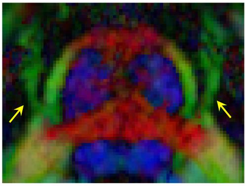
Diffusion tensor imaging (DTI) can provide markers of axonal micro‐structure of the trigeminal nerve (cranial nerve five [CNV]), which may be affected in trigeminal neuralgia (TN) and other disorders. Previous attempts to image CNV have used low spatial resolution DTI protocols designed for whole‐brain acquisition that are susceptible to errors from partial volume effects, particularly with adjacent cerebrospinal fluid (CSF). The purpose of this study was to develop a nerve‐specific DTI protocol in healthy subjects that provides more accurate CNV tractography and diffusion quantification than whole‐brain protocols. Four DTI protocols were compared in five healthy individuals (age 22–45 years, three males) on a 3 T Siemens Prisma MRI scanner: two newly developed nerve‐specific high resolution (1.2 x 1.2 x 1.2 = 1.7 mm3) DTI protocols without (3.5 minutes) and with CSF suppression (fluid‐attenuated inversion recovery [FLAIR]; 7.5 minutes) with limited slice‐coverage, and two typical whole‐brain protocols with either isotropic (2 x 2 x 2 = 8 mm3) or thicker slice anisotropic (1.9 x 1.9 x 3 = 10.8 mm3) voxels. Deterministic tractography was used to identify the CNV and quantify bilateral fractional anisotropy (FA), and mean (MD), axial (AD) and radial diffusivity (RD). CNV volume was determined by manual tracing on T1‐weighted images. High spatial resolution nerve‐specific protocols yielded better delineation of CNV, with less distortions and blurring, and markedly different diffusion parameters (42% higher FA, 35% lower MD, 27% lower RD and 43% lower AD) compared with the two lower resolution whole‐brain protocols. The anisotropic whole‐brain protocol showed a positive correlation between CNV FA and volume. The high resolution nerve‐specific protocol with FLAIR yielded additional reductions in CNV AD and MD with a value of 1.0 x 10⁻³ mm2/s, approaching that expected for healthy young adult white matter. In conclusion, high resolution nerve‐specific DTI with FLAIR enhances the identification of CNV and provides more accurate quantification of diffusion compared with lower resolution whole‐brain approaches.
中文翻译:

高空间分辨率神经特异性 DTI 协议优于全脑 DTI 协议,用于对健康个体的三叉神经进行成像
弥散张量成像 (DTI) 可以提供三叉神经(第 5 颅神经 [CNV])轴突微结构的标记,这可能会影响三叉神经痛 (TN) 和其他疾病。以前对 CNV 进行成像的尝试使用了专为全脑采集而设计的低空间分辨率 DTI 协议,这些协议易受部分体积效应引起的错误影响,尤其是邻近脑脊液 (CSF)。本研究的目的是在健康受试者中开发一种神经特异性 DTI 协议,该协议提供比全脑协议更准确的 CNV 纤维束成像和扩散量化。在 3 T Siemens Prisma MRI 扫描仪上对五名健康个体(22-45 岁,三名男性)的四种 DTI 协议进行了比较:两种新开发的神经特异性高分辨率(1.2 x 1.2 x 1.2 = 1.7 mm 3) 没有(3.5 分钟)和有 CSF 抑制(流体衰减反转恢复 [FLAIR];7.5 分钟)且切片覆盖有限的 DTI 协议,以及两种具有各向同性(2 x 2 x 2 = 8 mm)的典型全脑协议3 ) 或更厚的切片各向异性 (1.9 x 1.9 x 3 = 10.8 mm 3) 体素。确定性纤维束成像用于识别 CNV 并量化双侧各向异性分数 (FA) 和平均值 (MD)、轴向 (AD) 和径向扩散率 (RD)。CNV 体积是通过在 T1 加权图像上手动追踪来确定的。与两者相比,高空间分辨率神经特异性方案产生了更好的 CNV 描绘,失真和模糊更少,扩散参数明显不同(FA 高 42%,MD 低 35%,RD 低 27%,AD 低 43%)分辨率全脑协议。各向异性全脑协议显示 CNV FA 与体积之间呈正相关。使用 FLAIR 的高分辨率神经特异性方案使 CNV AD 和 MD 进一步降低,值为 1.0 x 10 -3 mm 2/s,接近健康年轻成人白质的预期值。总之,与较低分辨率的全脑方法相比,具有 FLAIR 的高分辨率神经特异性 DTI 增强了 CNV 的识别并提供了更准确的扩散量化。
更新日期:2020-10-10
中文翻译:
高空间分辨率神经特异性 DTI 协议优于全脑 DTI 协议,用于对健康个体的三叉神经进行成像
弥散张量成像 (DTI) 可以提供三叉神经(第 5 颅神经 [CNV])轴突微结构的标记,这可能会影响三叉神经痛 (TN) 和其他疾病。以前对 CNV 进行成像的尝试使用了专为全脑采集而设计的低空间分辨率 DTI 协议,这些协议易受部分体积效应引起的错误影响,尤其是邻近脑脊液 (CSF)。本研究的目的是在健康受试者中开发一种神经特异性 DTI 协议,该协议提供比全脑协议更准确的 CNV 纤维束成像和扩散量化。在 3 T Siemens Prisma MRI 扫描仪上对五名健康个体(22-45 岁,三名男性)的四种 DTI 协议进行了比较:两种新开发的神经特异性高分辨率(1.2 x 1.2 x 1.2 = 1.7 mm 3) 没有(3.5 分钟)和有 CSF 抑制(流体衰减反转恢复 [FLAIR];7.5 分钟)且切片覆盖有限的 DTI 协议,以及两种具有各向同性(2 x 2 x 2 = 8 mm)的典型全脑协议3 ) 或更厚的切片各向异性 (1.9 x 1.9 x 3 = 10.8 mm 3) 体素。确定性纤维束成像用于识别 CNV 并量化双侧各向异性分数 (FA) 和平均值 (MD)、轴向 (AD) 和径向扩散率 (RD)。CNV 体积是通过在 T1 加权图像上手动追踪来确定的。与两者相比,高空间分辨率神经特异性方案产生了更好的 CNV 描绘,失真和模糊更少,扩散参数明显不同(FA 高 42%,MD 低 35%,RD 低 27%,AD 低 43%)分辨率全脑协议。各向异性全脑协议显示 CNV FA 与体积之间呈正相关。使用 FLAIR 的高分辨率神经特异性方案使 CNV AD 和 MD 进一步降低,值为 1.0 x 10 -3 mm 2/s,接近健康年轻成人白质的预期值。总之,与较低分辨率的全脑方法相比,具有 FLAIR 的高分辨率神经特异性 DTI 增强了 CNV 的识别并提供了更准确的扩散量化。


 京公网安备 11010802027423号