当前位置:
X-MOL 学术
›
Microsc. Res. Tech.
›
论文详情
Our official English website, www.x-mol.net, welcomes your
feedback! (Note: you will need to create a separate account there.)
Microscopic and spectroscopic characterization of an extraskeletal intranasal osteoma in a Gir cow
Microscopy Research and Technique ( IF 2.0 ) Pub Date : 2020-10-09 , DOI: 10.1002/jemt.23613 Vineet Kumar 1 , Foram A Asodiya 2 , Vivek K Singh 3 , Harsukh P Gajera 4
Microscopy Research and Technique ( IF 2.0 ) Pub Date : 2020-10-09 , DOI: 10.1002/jemt.23613 Vineet Kumar 1 , Foram A Asodiya 2 , Vivek K Singh 3 , Harsukh P Gajera 4
Affiliation
|
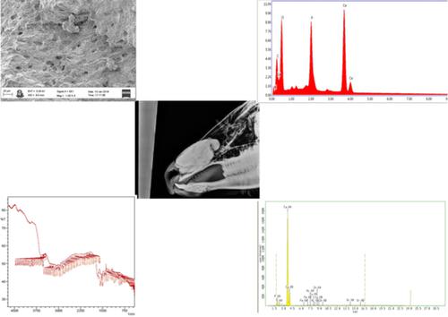
This is probably the first report characterizing an extraskeletal intranasal osteoma in a Gir cow through scanning electron microscopy and various spectroscopic techniques. Nasal obstruction in a 10‐year‐old Gir cow is investigated in this study. Skull radiograph demonstrated 174.12 mm × 81.97 mm sized well‐circumscribed radiodense mass within the left nasal passage. The intranasal mass was excised completely through a rhinotomy incision. Grossly, intranasal mass was nonhyperemic, rock‐hard, and calcified, 174.12 mm × 81.97 mm in size, and 650 g of weight. Excised intranasal mass was investigated through histopathologic, scanning electron microscopic (SEM), energy‐dispersive X‐ray (EDX) spectroscopic, X‐ray fluorescence (XRF) spectroscopic, microwave plasma‐atomic energy spectroscopic (MPAES), and Fourier‐transform infrared (FTIR) spectroscopic techniques. A native bone of age‐matched Gir cow, collected from a cadaver, was taken as a control. Microscopically, structures similar to cortical bone randomly coexisted with trabecular bone were observed. The EDX analysis of the intranasal mass indicated mean Ca/P weight ratio of 1.88, close to Ca/P weight ratio of the control. The XRF analysis revealed the presence of Ca, P, Sr, S, Zn, Cu, Fe, and Ni in the intranasal mass. Additionally, Mn was noted by MPAES analysis. Hence, the XRF and MPAES analyses confirmed a similar elemental composition of the intranasal mass and control. FTIR spectroscopic study confirmed the presence of inorganic ν1, ν3 PO43−, OH− in addition to organic collagen amide A, amide B, amide I, amide II, and amide III chemical functional groups in the intranasal mass. These findings of the intranasal mass were consistent with an osteoma having similar elemental and molecular compositions with the native bone.
中文翻译:

吉尔奶牛骨骼外鼻内骨瘤的显微和光谱表征
这可能是第一份通过扫描电子显微镜和各种光谱技术表征 Gir 奶牛骨骼外鼻内骨瘤的报告。本研究调查了 10 岁 Gir 奶牛的鼻塞。头骨 X 光片显示左侧鼻道内有 174.12 mm × 81.97 mm 大小的边界清楚的放射性密度肿块。通过鼻切开切口完全切除鼻内肿块。大体上,鼻内肿块是非充血性的、坚硬的、钙化的,大小为 174.12 mm × 81.97 mm,重量为 650 g。通过组织病理学、扫描电子显微镜 (SEM)、能量色散 X 射线 (EDX) 光谱、X 射线荧光 (XRF) 光谱、微波等离子体原子能谱 (MPAES)、和傅里叶变换红外(FTIR)光谱技术。从尸体中收集的年龄匹配的 Gir 牛的原生骨骼作为对照。显微镜下观察到类似皮质骨的结构与小梁骨随机共存。鼻内物质的 EDX 分析表明平均 Ca/P 重量比为 1.88,接近对照的 Ca/P 重量比。XRF 分析显示鼻内肿块中存在 Ca、P、Sr、S、Zn、Cu、Fe 和 Ni。此外,通过 MPAES 分析记录了 Mn。因此,XRF 和 MPAES 分析证实了鼻内物质和对照的相似元素组成。FTIR 光谱研究证实了无机 ν 的存在 观察到类似于皮质骨的结构与小梁骨随机共存。鼻内物质的 EDX 分析表明平均 Ca/P 重量比为 1.88,接近对照的 Ca/P 重量比。XRF 分析显示鼻内肿块中存在 Ca、P、Sr、S、Zn、Cu、Fe 和 Ni。此外,通过 MPAES 分析记录了 Mn。因此,XRF 和 MPAES 分析证实了鼻内物质和对照的相似元素组成。FTIR 光谱研究证实了无机 ν 的存在 观察到类似于皮质骨的结构与小梁骨随机共存。鼻内物质的 EDX 分析表明平均 Ca/P 重量比为 1.88,接近对照的 Ca/P 重量比。XRF 分析显示鼻内肿块中存在 Ca、P、Sr、S、Zn、Cu、Fe 和 Ni。此外,通过 MPAES 分析记录了 Mn。因此,XRF 和 MPAES 分析证实了鼻内物质和对照的相似元素组成。FTIR 光谱研究证实了无机 ν 的存在 因此,XRF 和 MPAES 分析证实了鼻内物质和对照的相似元素组成。FTIR 光谱研究证实了无机 ν 的存在 因此,XRF 和 MPAES 分析证实了鼻内物质和对照的相似元素组成。FTIR 光谱研究证实了无机 ν 的存在1, ν 3 PO 4 3- , OH -除了鼻内物质中的有机胶原酰胺A、酰胺B、酰胺I、酰胺II和酰胺III化学官能团。鼻内肿块的这些发现与骨瘤一致,该骨瘤具有与天然骨相似的元素和分子组成。
更新日期:2020-10-09
中文翻译:
吉尔奶牛骨骼外鼻内骨瘤的显微和光谱表征
这可能是第一份通过扫描电子显微镜和各种光谱技术表征 Gir 奶牛骨骼外鼻内骨瘤的报告。本研究调查了 10 岁 Gir 奶牛的鼻塞。头骨 X 光片显示左侧鼻道内有 174.12 mm × 81.97 mm 大小的边界清楚的放射性密度肿块。通过鼻切开切口完全切除鼻内肿块。大体上,鼻内肿块是非充血性的、坚硬的、钙化的,大小为 174.12 mm × 81.97 mm,重量为 650 g。通过组织病理学、扫描电子显微镜 (SEM)、能量色散 X 射线 (EDX) 光谱、X 射线荧光 (XRF) 光谱、微波等离子体原子能谱 (MPAES)、和傅里叶变换红外(FTIR)光谱技术。从尸体中收集的年龄匹配的 Gir 牛的原生骨骼作为对照。显微镜下观察到类似皮质骨的结构与小梁骨随机共存。鼻内物质的 EDX 分析表明平均 Ca/P 重量比为 1.88,接近对照的 Ca/P 重量比。XRF 分析显示鼻内肿块中存在 Ca、P、Sr、S、Zn、Cu、Fe 和 Ni。此外,通过 MPAES 分析记录了 Mn。因此,XRF 和 MPAES 分析证实了鼻内物质和对照的相似元素组成。FTIR 光谱研究证实了无机 ν 的存在 观察到类似于皮质骨的结构与小梁骨随机共存。鼻内物质的 EDX 分析表明平均 Ca/P 重量比为 1.88,接近对照的 Ca/P 重量比。XRF 分析显示鼻内肿块中存在 Ca、P、Sr、S、Zn、Cu、Fe 和 Ni。此外,通过 MPAES 分析记录了 Mn。因此,XRF 和 MPAES 分析证实了鼻内物质和对照的相似元素组成。FTIR 光谱研究证实了无机 ν 的存在 观察到类似于皮质骨的结构与小梁骨随机共存。鼻内物质的 EDX 分析表明平均 Ca/P 重量比为 1.88,接近对照的 Ca/P 重量比。XRF 分析显示鼻内肿块中存在 Ca、P、Sr、S、Zn、Cu、Fe 和 Ni。此外,通过 MPAES 分析记录了 Mn。因此,XRF 和 MPAES 分析证实了鼻内物质和对照的相似元素组成。FTIR 光谱研究证实了无机 ν 的存在 因此,XRF 和 MPAES 分析证实了鼻内物质和对照的相似元素组成。FTIR 光谱研究证实了无机 ν 的存在 因此,XRF 和 MPAES 分析证实了鼻内物质和对照的相似元素组成。FTIR 光谱研究证实了无机 ν 的存在1, ν 3 PO 4 3- , OH -除了鼻内物质中的有机胶原酰胺A、酰胺B、酰胺I、酰胺II和酰胺III化学官能团。鼻内肿块的这些发现与骨瘤一致,该骨瘤具有与天然骨相似的元素和分子组成。


 京公网安备 11010802027423号