当前位置:
X-MOL 学术
›
J. Cell. Physiol.
›
论文详情
Our official English website, www.x-mol.net, welcomes your
feedback! (Note: you will need to create a separate account there.)
IL‐1/IL‐1R signaling induced by all‐trans‐retinal contributes to complement alternative pathway activation in retinal pigment epithelium
Journal of Cellular Physiology ( IF 4.5 ) Pub Date : 2020-10-09 , DOI: 10.1002/jcp.30103 Xinxuan Cheng 1, 2, 3 , Danxue He 1, 2, 3 , Chunyan Liao 1, 2, 3 , Sijie Lin 1, 2, 3 , Liying Tang 1, 2, 3 , Yuan-Liang Wang 4, 5 , Jiaoyue Hu 1, 2, 3 , Wei Li 1, 2, 3 , Zuguo Liu 1, 2, 3 , Yalin Wu 1, 2, 3 , Yi Liao 1, 2, 3
Journal of Cellular Physiology ( IF 4.5 ) Pub Date : 2020-10-09 , DOI: 10.1002/jcp.30103 Xinxuan Cheng 1, 2, 3 , Danxue He 1, 2, 3 , Chunyan Liao 1, 2, 3 , Sijie Lin 1, 2, 3 , Liying Tang 1, 2, 3 , Yuan-Liang Wang 4, 5 , Jiaoyue Hu 1, 2, 3 , Wei Li 1, 2, 3 , Zuguo Liu 1, 2, 3 , Yalin Wu 1, 2, 3 , Yi Liao 1, 2, 3
Affiliation
|
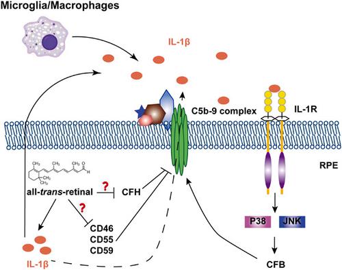
The underlying mechanisms of complement activation in Stargardt disease type 1 (STGD1) and age‐related macular degeneration (AMD) are not fully understood. Overaccumulation of all‐trans‐retinal (atRAL) has been proposed as the pathogenic factor in both diseases. By incubating retinal pigment epithelium (RPE) cells with atRAL, we showed that C5b‐9 membrane attack complexes (MACs) were generated mainly through complement alternative pathway. An increase in complement factor B (CFB) expression as well as downregulation of complement regulatory proteins CD46, CD55, CD59, and CFH were observed in RPE cells after atRAL treatment. Furthermore, interleukin‐1β production was provoked in both atRAL‐treated RPE cells and microglia/macrophages. Coincubation of RPE cells with interleukin‐1 receptor antagonist (IL1Ra) and atRAL ameliorated complement activation and downregulated CFB expression by attenuating both p38 and c‐Jun N‐terminal kinase (JNK) signaling pathways. Our findings demonstrate that atRAL induces an autocrine/paracrine IL‐1/IL‐1R signaling to promote complement alternative pathway activation in RPE cells and provide a novel perspective on the pathomechanism of macular degeneration.
中文翻译:

全反式视网膜诱导的 IL-1/IL-1R 信号传导有助于视网膜色素上皮细胞中的补体旁路激活
Stargardt 病 1 型(STGD1)和年龄相关性黄斑变性(AMD)中补体激活的潜在机制尚不完全清楚。全反式的过度积累视网膜(atRAL)被认为是这两种疾病的致病因素。通过将视网膜色素上皮 (RPE) 细胞与 atRAL 一起孵育,我们发现 C5b-9 膜攻击复合物 (MAC) 主要通过补体替代途径产生。在 atRAL 处理后,在 RPE 细胞中观察到补体因子 B (CFB) 表达增加以及补体调节蛋白 CD46、CD55、CD59 和 CFH 的下调。此外,在经 atRAL 处理的 RPE 细胞和小胶质细胞/巨噬细胞中均激发了白细胞介素-1β 的产生。RPE 细胞与白细胞介素 1 受体拮抗剂 (IL1Ra) 和 atRAL 的共孵育通过减弱 p38 和 c-Jun N 末端激酶 (JNK) 信号通路来改善补体激活并下调 CFB 表达。
更新日期:2020-10-09
中文翻译:
全反式视网膜诱导的 IL-1/IL-1R 信号传导有助于视网膜色素上皮细胞中的补体旁路激活
Stargardt 病 1 型(STGD1)和年龄相关性黄斑变性(AMD)中补体激活的潜在机制尚不完全清楚。全反式的过度积累视网膜(atRAL)被认为是这两种疾病的致病因素。通过将视网膜色素上皮 (RPE) 细胞与 atRAL 一起孵育,我们发现 C5b-9 膜攻击复合物 (MAC) 主要通过补体替代途径产生。在 atRAL 处理后,在 RPE 细胞中观察到补体因子 B (CFB) 表达增加以及补体调节蛋白 CD46、CD55、CD59 和 CFH 的下调。此外,在经 atRAL 处理的 RPE 细胞和小胶质细胞/巨噬细胞中均激发了白细胞介素-1β 的产生。RPE 细胞与白细胞介素 1 受体拮抗剂 (IL1Ra) 和 atRAL 的共孵育通过减弱 p38 和 c-Jun N 末端激酶 (JNK) 信号通路来改善补体激活并下调 CFB 表达。


 京公网安备 11010802027423号