Computer Methods and Programs in Biomedicine ( IF 4.9 ) Pub Date : 2020-10-08 , DOI: 10.1016/j.cmpb.2020.105791 Yanhui Guo , Guo-Qing Du , Wen-Qian Shen , Chunlai Du , Pei-Na He , Siuly Siuly
|
Purpose
Heart disease is one of the leading causes of death. Among patients with cardiovascular diseases, myocardial infarction (MI) is the main reason. Precise and timely identification of MI is significant for early treatment. Myocardial contrast echocardiography (MCE) is widely used for the detection of MI in clinic practice. However, existing clinical exam using MCE is subjective and highly operator dependent and time-consuming. Hence an automatic computer-aided MI detection in MCE is necessary to improve the diagnosis performance and decrease the workload of clinicians.
Methods
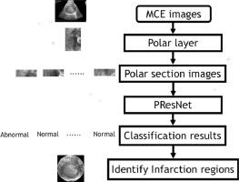
In this study, a novel deep learning model, polar residual network (PResNet) is proposed to identify MI regions in MCE images which design a polar layer considering the ring shape of the myocardium. MCE images are fed into the PResNet and a newly defined polar layer is used to describe the myocardium with a ring shape. The whole polar images are evenly divided into several subsections and a residual network is improved to classify the subsection into normal and abnormal categories. Finally, the detection results are mapped back to the original image to illustrate the infarction regions’ locations for the further process.
Results
To evaluate the proposed PResNet, a dataset is constructed via performing MCE on five mice, which underwent the left anterior descending artery ligation and receive erythropoietin or saline injection, and the area variation fraction is manually annotated by an experienced expert as golden standards. The results demonstrate that the proposed PResNet model accomplishes high classification precisions with 99.6% and 98.7%, and 0.999 and 0.996 of AUC (area under the receiver operator curve) values on two different testing sets, respectively. Results suggest that the proposed model could enable accurate infarct detection and diagnosis of the MCE images.
Conclusion
Those efficiency gains highlight the powerful ability to describe and interpret the MCE images using the polar layer and residual network. The proposed PResNet might aid the clinicians in fast and accurate assessing the infarcted myocardium on MCE.
中文翻译:
基于极性残差网络的对比超声心动图自动检测心肌梗塞
目的
心脏病是导致死亡的主要原因之一。在患有心血管疾病的患者中,心肌梗塞(MI)是主要原因。准确,及时地识别心肌梗死对于早期治疗很重要。心肌对比超声心动图(MCE)在临床实践中广泛用于检测MI。但是,使用MCE进行的现有临床检查是主观的,并且高度依赖操作员且耗时。因此,在MCE中自动进行计算机辅助MI检测对于提高诊断性能和减少临床医生的工作量是必要的。
方法
在这项研究中,提出了一种新颖的深度学习模型,极地残差网络(PResNet)来识别MCE图像中的MI区域,该区域设计了考虑心肌环形状的极地层。MCE图像被馈入PResNet,并使用新定义的极性层来描述具有环形形状的心肌。将整个极地图像平均分为几个子部分,并对残差网络进行改进以将子部分分为正常和异常类别。最后,将检测结果映射回原始图像,以说明梗塞区域的位置,以便进行进一步处理。
结果
为了评估拟议的PResNet,通过对五只小鼠进行MCE来构建数据集,对它们进行左前降支结扎并接受促红细胞生成素或生理盐水注射,并且由经验丰富的专家手动将面积变化分数注释为黄金标准。结果表明,所提出的PResNet模型在两个不同的测试集上分别实现了99.6%和98.7%的AUC值(接收者操作员曲线下的面积)分别为99.6%和98.7%的高分类精度。结果表明,提出的模型可以实现MCE图像的准确梗塞检测和诊断。
结论
这些效率的提高突显了使用极地层和残留网络描述和解释MCE图像的强大能力。拟议的PResNet可能有助于临床医生快速准确地评估MCE上的梗塞心肌。


 京公网安备 11010802027423号