Computers in Biology and Medicine ( IF 7.0 ) Pub Date : 2020-09-30 , DOI: 10.1016/j.compbiomed.2020.104019 Vy Bui 1 , Li-Yueh Hsu 2 , Sujata M Shanbhag 3 , Loc Tran 4 , W Patricia Bandettini 3 , Lin-Ching Chang 4 , Marcus Y Chen 3
|
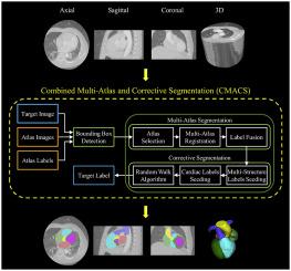
Multi-atlas based segmentation is an effective technique that transforms a representative set of atlas images and labels into a target image for structural segmentation. However, a significant limitation of this approach relates to the fact that the atlas and the target images need to be similar in volume orientation, coverage, or acquisition protocols in order to prevent image misregistration and avoid segmentation fault. In this study, we aim to evaluate the impact of using a heterogeneous Computed Tomography Angiography (CTA) dataset on the performance of a multi-atlas cardiac structure segmentation framework. We propose a generalized technique based upon using the Simple Linear Iterative Clustering (SLIC) supervoxel method to detect a bounding box region enclosing the heart before subsequent cardiac structure segmentation. This technique facilitates our framework to process CTA datasets acquired from distinct imaging protocols and to improve its segmentation accuracy and speed. In a four-way cross comparison based on 60 CTA studies from our institution and 60 CTA datasets from the Multi-Modality Whole Heart Segmentation MICCAI challenge, we show that the proposed framework performs well in segmenting seven different cardiac structures based upon interchangeable atlas and target datasets acquired from different imaging settings. For the overall results, our automated segmentation framework attains a median Dice, mean distance, and Hausdorff distance of 0.88, 1.5 mm, and 9.69 mm over the entire datasets. The average processing time was 1.55 minutes for both datasets. Furthermore, this study shows that it is feasible to exploit heterogenous datasets from different imaging protocols and institutions for accurate multi-atlas cardiac structure segmentation.
中文翻译:
改进计算机断层扫描血管造影的多图谱心脏结构分割:基于异构数据集的性能评估
基于多图谱的分割是一种有效的技术,它将一组代表性的图谱图像和标签转换为目标图像以进行结构分割。然而,这种方法的一个重大限制与以下事实有关:图集和目标图像需要在体积方向、覆盖范围或采集协议方面相似,以防止图像重合并避免分割错误。在本研究中,我们的目的是评估使用异构计算机断层扫描血管造影(CTA)数据集对多图谱心脏结构分割框架性能的影响。我们提出了一种基于使用简单线性迭代聚类(SLIC)超体素方法的通用技术,在随后的心脏结构分割之前检测包围心脏的边界框区域。这项技术有助于我们的框架处理从不同成像协议获取的 CTA 数据集,并提高其分割准确性和速度。在基于我们机构的 60 个 CTA 研究和来自多模态全心脏分割 MICCAI 挑战的 60 个 CTA 数据集的四向交叉比较中,我们表明,所提出的框架在基于可互换的图集和目标分割七种不同的心脏结构方面表现良好从不同成像设置获取的数据集。对于总体结果,我们的自动分割框架在整个数据集上获得了 0.88、1.5 毫米和 9.69 毫米的中值 Dice、平均距离和豪斯多夫距离。两个数据集的平均处理时间均为 1.55 分钟。此外,这项研究表明,利用来自不同成像协议和机构的异构数据集进行精确的多图谱心脏结构分割是可行的。


 京公网安备 11010802027423号