NeuroImage: Clinical ( IF 3.4 ) Pub Date : 2020-09-28 , DOI: 10.1016/j.nicl.2020.102447 Natalie R Boonzaier 1 , Patrick W Hales 1 , Felice D'Arco 2 , Bronwen C Walters 2 , Ramneek Kaur 1 , Kshitij Mankad 2 , Jessica Cooper 2 , Alki Liasis 3 , Victoria Smith 2 , Patricia O'Hare 2 , Darren Hargrave 4 , Christopher A Clark 1
|
Background
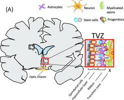
The subventricular zone of the third ventricle (TVZ) is a germinal stem cell niche, identified as the possible location of optic pathway glioma (OPG) cell origin. Paediatric OPGs are predominantly diagnosed as low-grade astrocytomas, which are either sporadic or are associated with neurofibromatosis type-1 (NF1). These tumours often cause a significant impairment to visual acuity (VA). Infiltrative/invasive tumour activity is associated with increased apparent diffusion coefficient (ADC) and cerebral blood flow (CBF). This study aimed to determine whether TVZ imaging features differed between sporadic-OPG, NF1-OPG and controls, and whether the ADC and CBF profile at the germinal stem cell niche (the TVZ) correlated with the primary outcome of VA.
Methods
ADC and CBF MRI data were acquired from 30 paediatric OPG patients (median age 6 years; range 8 months–17 years), along with VA measurements, during clinical surveillance of their tumour. Values for mean ADC and maximum CBF were measured at the TVZ, and normalized to normal-appearing grey matter. These values were compared between the two OPG groups and the healthy control subjects, and multivariate linear regression was used to test the linear association between these values and patient’s VA.
Results
In the TVZ, normalized mean ADC was higher in NF1-associated OPG patients (N = 15), compared to both sporadic OPG patients (N = 15; p = 0.010) and healthy controls (N = 14; p < 0.001). In the same region, normalized maximum CBF was higher in sporadic OPG patients compared to both NF1-OPG patients (p = 0.016) and healthy controls (p < 0.001). In sporadic OPG patients only, normalized mean ADC in the TVZ was significantly correlated with visual acuity (R2 = 0.41, p = 0.019). No significant correlations were found between TVZ CBF and ADC values and visual acuity in the NF1-associated OPG patients.
Conclusion
Quantitative MRI detects TVZ abnormalities in both sporadic and NF1-OPG patients, and identifies TVZ features that differentiate the two. TVZ features may be useful MRI markers of interest in future predictive studies involving sporadic OPG.
中文翻译:
定量MRI显示1型神经纤维瘤病和散发性儿科视神经胶质瘤的第三脑室下区异常
背景
第三脑室(TVZ)的脑室下区是生发的干细胞小生境,被确定为视神经胶质瘤(OPG)细胞起源的可能位置。儿科OPG主要被诊断为低度星形细胞瘤,它们是散发性的或与1型神经纤维瘤病(NF1)相关。这些肿瘤通常会严重损害视力(VA)。浸润/浸润性肿瘤活动与表观扩散系数(ADC)和脑血流量(CBF)增加相关。这项研究旨在确定散发性OPG,NF1-OPG和对照之间TVZ成像特征是否不同,以及生发干细胞生态位(TVZ)的ADC和CBF谱是否与VA的主要结局相关。
方法
从30名儿科OPG患者(中位年龄6岁;范围8个月至17岁)中获得ADC和CBF MRI数据,并在其临床肿瘤监测期间进行VA测量。在TVZ上测量平均ADC和最大CBF的值,并将其标准化为正常显示的灰质。在两个OPG组和健康对照组之间比较这些值,并使用多元线性回归测试这些值与患者VA之间的线性关联。
结果
在TVZ中,与散发性OPG患者(N = 15; p = 0.010)和健康对照组(N = 14; p <0.001)相比,与NF1相关的OPG患者(N = 15)的标准化ADC均高。在同一地区,散发性OPG患者的正常最大CBF高于NF1-OPG患者(p = 0.016)和健康对照组(p <0.001)。仅在零星的OPG患者中,TVZ中的归一化平均ADC与视敏度显着相关(R 2 = 0.41,p = 0.019)。在与NF1相关的OPG患者中,TVZ CBF和ADC值与视敏度之间没有发现显着相关性。
结论
定量MRI可检测散发性和NF1-OPG患者的TVZ异常,并识别出区分两者的TVZ功能。TVZ功能可能是涉及散发性OPG的未来预测研究中感兴趣的有用MRI标记。


 京公网安备 11010802027423号