当前位置:
X-MOL 学术
›
Biosyst. Eng.
›
论文详情
Our official English website, www.x-mol.net, welcomes your
feedback! (Note: you will need to create a separate account there.)
Micro-CT imaging of tomato seeds: Predictive potential of 3D morphometry on germination
Biosystems Engineering ( IF 4.4 ) Pub Date : 2020-12-01 , DOI: 10.1016/j.biosystemseng.2020.09.003 Laura Gargiulo , Cristina Leonarduzzi , Giacomo Mele
Biosystems Engineering ( IF 4.4 ) Pub Date : 2020-12-01 , DOI: 10.1016/j.biosystemseng.2020.09.003 Laura Gargiulo , Cristina Leonarduzzi , Giacomo Mele
|
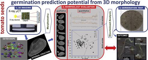
The relationship between seed physical characteristics and seed quality is widely investigated by using X-ray based imaging techniques. Recently the use of X-ray micro-tomography (micro-CT) is increasingly used for more accurate characterisation of the internal seed morphology. In this work a germination test was carried out along with the morphometric characterisation of tomato seed internal structure by means of X-ray micro-CT and 3D image analysis. The aim was to accurately evaluate the predictive potential of internal seed 3D morphology for germination outcomes. The visual assessment allowed the relationship between specific internal seed abnormalities and the different germination outcomes to be demonstrated experimentally. Univariate analysis of morphometric seed traits allowed 3D free space % and Sauter diameter, among the most discriminant parameters, to be identified as the most effective for the prediction of germination outcomes. Discriminant Analysis (DA) of 3D morphometric dataset correctly classified 96.3% of normal seedlings, 83.3% of ungerminated seeds and 63.6% of abnormal seedlings, providing a high overall prediction potential of 91.9%. The above analyses have also been performed referring to the germination at 5 days after sowing. As a side effect of the applied technique, an increase of abnormal seedlings was observed at increasing X-ray exposure level. Overall, X-ray micro-CT coupled with DA of internal morphometric traits has proved to be an effective tool to investigate the relationship between tomato seed 3D morphology and seed physiology, although attention has to be paid to possible consequences of X-ray exposure.
中文翻译:

番茄种子的显微 CT 成像:3D 形态测量学对萌发的预测潜力
使用基于 X 射线的成像技术广泛研究了种子物理特性和种子质量之间的关系。最近,X 射线显微断层扫描 (micro-CT) 越来越多地用于更准确地表征内部种子形态。在这项工作中,通过 X 射线显微 CT 和 3D 图像分析,进行了发芽测试以及番茄种子内部结构的形态测量表征。目的是准确评估内部种子 3D 形态对发芽结果的预测潜力。视觉评估允许通过实验证明特定内部种子异常与不同发芽结果之间的关系。形态计量种子性状的单变量分析允许 3D 自由空间百分比和索特直径,在最具辨别力的参数中,被确定为预测发芽结果最有效的参数。3D 形态测量数据集的判别分析 (DA) 正确分类了 96.3% 的正常幼苗、83.3% 的未发芽种子和 63.6% 的异常幼苗,提供了 91.9% 的高总体预测潜力。上述分析也针对播种后 5 天的发芽情况进行了分析。作为应用技术的一个副作用,在增加 X 射线暴露水平时观察到异常幼苗的增加。总体而言,X 射线显微 CT 加上内部形态特征的 DA 已被证明是研究番茄种子 3D 形态与种子生理学之间关系的有效工具,尽管必须注意 X 射线暴露的可能后果。被确定为预测发芽结果最有效的方法。3D 形态测量数据集的判别分析 (DA) 正确分类了 96.3% 的正常幼苗、83.3% 的未发芽种子和 63.6% 的异常幼苗,提供了 91.9% 的高总体预测潜力。上述分析也针对播种后 5 天的发芽情况进行了分析。作为应用技术的一个副作用,在增加 X 射线暴露水平时观察到异常幼苗的增加。总体而言,X 射线显微 CT 加上内部形态特征的 DA 已被证明是研究番茄种子 3D 形态与种子生理学之间关系的有效工具,尽管必须注意 X 射线暴露的可能后果。被确定为预测发芽结果最有效的方法。3D 形态测量数据集的判别分析 (DA) 正确分类了 96.3% 的正常幼苗、83.3% 的未发芽种子和 63.6% 的异常幼苗,提供了 91.9% 的高总体预测潜力。上述分析也针对播种后 5 天的发芽情况进行了分析。作为应用技术的一个副作用,在增加 X 射线暴露水平时观察到异常幼苗的增加。总体而言,X 射线显微 CT 加上内部形态特征的 DA 已被证明是研究番茄种子 3D 形态与种子生理学之间关系的有效工具,尽管必须注意 X 射线暴露的可能后果。
更新日期:2020-12-01
中文翻译:
番茄种子的显微 CT 成像:3D 形态测量学对萌发的预测潜力
使用基于 X 射线的成像技术广泛研究了种子物理特性和种子质量之间的关系。最近,X 射线显微断层扫描 (micro-CT) 越来越多地用于更准确地表征内部种子形态。在这项工作中,通过 X 射线显微 CT 和 3D 图像分析,进行了发芽测试以及番茄种子内部结构的形态测量表征。目的是准确评估内部种子 3D 形态对发芽结果的预测潜力。视觉评估允许通过实验证明特定内部种子异常与不同发芽结果之间的关系。形态计量种子性状的单变量分析允许 3D 自由空间百分比和索特直径,在最具辨别力的参数中,被确定为预测发芽结果最有效的参数。3D 形态测量数据集的判别分析 (DA) 正确分类了 96.3% 的正常幼苗、83.3% 的未发芽种子和 63.6% 的异常幼苗,提供了 91.9% 的高总体预测潜力。上述分析也针对播种后 5 天的发芽情况进行了分析。作为应用技术的一个副作用,在增加 X 射线暴露水平时观察到异常幼苗的增加。总体而言,X 射线显微 CT 加上内部形态特征的 DA 已被证明是研究番茄种子 3D 形态与种子生理学之间关系的有效工具,尽管必须注意 X 射线暴露的可能后果。被确定为预测发芽结果最有效的方法。3D 形态测量数据集的判别分析 (DA) 正确分类了 96.3% 的正常幼苗、83.3% 的未发芽种子和 63.6% 的异常幼苗,提供了 91.9% 的高总体预测潜力。上述分析也针对播种后 5 天的发芽情况进行了分析。作为应用技术的一个副作用,在增加 X 射线暴露水平时观察到异常幼苗的增加。总体而言,X 射线显微 CT 加上内部形态特征的 DA 已被证明是研究番茄种子 3D 形态与种子生理学之间关系的有效工具,尽管必须注意 X 射线暴露的可能后果。被确定为预测发芽结果最有效的方法。3D 形态测量数据集的判别分析 (DA) 正确分类了 96.3% 的正常幼苗、83.3% 的未发芽种子和 63.6% 的异常幼苗,提供了 91.9% 的高总体预测潜力。上述分析也针对播种后 5 天的发芽情况进行了分析。作为应用技术的一个副作用,在增加 X 射线暴露水平时观察到异常幼苗的增加。总体而言,X 射线显微 CT 加上内部形态特征的 DA 已被证明是研究番茄种子 3D 形态与种子生理学之间关系的有效工具,尽管必须注意 X 射线暴露的可能后果。


 京公网安备 11010802027423号