当前位置:
X-MOL 学术
›
Microsc. Res. Tech.
›
论文详情
Our official English website, www.x-mol.net, welcomes your
feedback! (Note: you will need to create a separate account there.)
Anatomy and histology of reproductive system of adult male mint leaf beetle Chrysolina herbacea (Duftschmid, 1825) (Coleoptera: Chrysomelidae)
Microscopy Research and Technique ( IF 2.0 ) Pub Date : 2020-09-25 , DOI: 10.1002/jemt.23607 Nurcan Özyurt Koçakoğlu 1 , Selami Candan 1 , Mustafa Güllü 2
Microscopy Research and Technique ( IF 2.0 ) Pub Date : 2020-09-25 , DOI: 10.1002/jemt.23607 Nurcan Özyurt Koçakoğlu 1 , Selami Candan 1 , Mustafa Güllü 2
Affiliation
|
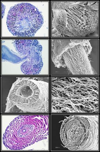
In this study, anatomy and histology of the male reproductive system of Chrysolina herbacea (Duftschmid 1825) (Coleoptera: Chrysomelidae) are described and illustrated by using light and scanning electron microscopies. Data from the male gonad of this species provide more extended and precise knowledge regarding the histoanatomical structure of the reproductive system in Chrysomelidae. The male reproductive system in C. herbacea consists of paired bilobed testes, two paired vas efferentia, paired seminal vesicles, paired vas deferentia, paired tubular accessory glands, a ductus ejaculatorius, and an aedeagus. Each testis consists of 40 follicles enveloped in a yellow pigmented sheath. In the light and scanning electron microscope examinations of male reproductive system of C. herbacea, different spermatogenesis stages (spermatocytes, spermatids, and spermatozoa) are found. Each testes follicle joins with the vas efferens surrounded by monolayered cubic epithelium with oval nuclei. Epithelial cells are covered from the outside with a thin layer of muscle and sheath. Vas efferens connect to vas deferens that may have an enlarged and differentiated region = seminal vesicle. Mature spermatozoa are seen in the lumen of the vas efferens, seminal vesicle, and vas deferens. There is a pair of accessory glands with a convoluted appearance in the tubular structure around the vas deferens. The vas deferens is a straight tube which leads into the proximal end of the ductus ejaculatorius. Ductus ejaculatorius wall is surrounded by intima, monolayer epithelium, and a thick muscle layer with many nuclei. The distal section of the ductus ejaculatorius is housed within the aedeagus.
中文翻译:

成年雄性薄荷叶甲虫 Chrysolina herbacea (Duftschmid, 1825) 生殖系统的解剖学和组织学(鞘翅目:叶甲科)
在这项研究中,通过使用光学和扫描电子显微镜描述和说明了Chrysolina herbacea(Duftschmid 1825)(鞘翅目:叶甲科)雄性生殖系统的解剖学和组织学。来自该物种雄性性腺的数据提供了有关叶甲科生殖系统组织解剖结构的更广泛和精确的知识。草本植物的雄性生殖系统由成对的双叶睾丸、两对输精管、成对的精囊、成对的输精管、成对的管状附属腺、射精管和阳茎组成。每个睾丸由 40 个被黄色鞘包裹的卵泡组成。在光镜和扫描电镜检查中,对草本植物雄性生殖系统进行了观察,发现了不同的精子发生阶段(精母细胞、精子细胞和精子)。每个睾丸滤泡与输精管相连,输精管被具有椭圆形核的单层立方上皮包围。上皮细胞从外部覆盖有一层薄薄的肌肉和鞘。输精管与输精管相连,输精管可能具有扩大和分化的区域=精囊。成熟精子可见于输精管、精囊和输精管的管腔中。输精管周围的管状结构中有一对外观复杂的副腺。输精管是通向射精管近端的直管。射精管壁被内膜、单层上皮和含有许多细胞核的厚肌肉层包围。射精管的远端部分位于阳茎内。
更新日期:2020-09-25
中文翻译:
成年雄性薄荷叶甲虫 Chrysolina herbacea (Duftschmid, 1825) 生殖系统的解剖学和组织学(鞘翅目:叶甲科)
在这项研究中,通过使用光学和扫描电子显微镜描述和说明了Chrysolina herbacea(Duftschmid 1825)(鞘翅目:叶甲科)雄性生殖系统的解剖学和组织学。来自该物种雄性性腺的数据提供了有关叶甲科生殖系统组织解剖结构的更广泛和精确的知识。草本植物的雄性生殖系统由成对的双叶睾丸、两对输精管、成对的精囊、成对的输精管、成对的管状附属腺、射精管和阳茎组成。每个睾丸由 40 个被黄色鞘包裹的卵泡组成。在光镜和扫描电镜检查中,对草本植物雄性生殖系统进行了观察,发现了不同的精子发生阶段(精母细胞、精子细胞和精子)。每个睾丸滤泡与输精管相连,输精管被具有椭圆形核的单层立方上皮包围。上皮细胞从外部覆盖有一层薄薄的肌肉和鞘。输精管与输精管相连,输精管可能具有扩大和分化的区域=精囊。成熟精子可见于输精管、精囊和输精管的管腔中。输精管周围的管状结构中有一对外观复杂的副腺。输精管是通向射精管近端的直管。射精管壁被内膜、单层上皮和含有许多细胞核的厚肌肉层包围。射精管的远端部分位于阳茎内。


 京公网安备 11010802027423号