当前位置:
X-MOL 学术
›
J. Raman Spectrosc.
›
论文详情
Our official English website, www.x-mol.net, welcomes your
feedback! (Note: you will need to create a separate account there.)
Bone grafted with β‐TCP granules in the rabbit: A microcomputed tomographic, histologic, Raman microspectrometric, and Raman imaging study
Journal of Raman Spectroscopy ( IF 2.4 ) Pub Date : 2020-09-25 , DOI: 10.1002/jrs.6000 Eric Aguado 1, 2 , Eric Goyenvalle 1, 2 , Claude Guintard 1, 2 , Daniel Chappard 2
Journal of Raman Spectroscopy ( IF 2.4 ) Pub Date : 2020-09-25 , DOI: 10.1002/jrs.6000 Eric Aguado 1, 2 , Eric Goyenvalle 1, 2 , Claude Guintard 1, 2 , Daniel Chappard 2
Affiliation
|
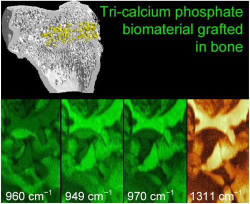
Bone grafting with synthetic substitutes is now the preferred technique in maxillofacial surgery. The biocompatibility of biomaterials such as beta‐tricalcium phosphate (β‐TCP) necessitates animal experimentation. Here, six rabbits received β‐TCP granules in a hole drilled in the femoral condyle and were compared with ungrafted holes with a similar critical size defect on the control‐lateral side. Microcomputed tomography confirmed that new bone trabeculae had invaded the grafted zone after 28 days, whereas only minimal remodeling had occurred at the margins of the hole in control rabbits. Histological sections, after polymethylmethacrylate (pMMA) embedding, evidenced a direct apposition of bone onto the granules. In some areas, osteoid tissue remained unmineralized, sandwiched between the biomaterial and fully calcified bone. Raman analysis was used to characterize the differences between the calcium/phosphate biomaterial and the bone matrix containing hydroxyapatite. Luminescence of β‐TCP was evidenced with a 785‐nm laser used for biological analysis but not with a 532‐nm laser. The ν1 phosphate allowed a clear differentiation of bone and the biomaterial as the subpeaks were not similar (960 cm−1 for bone, 949 and 970 cm−1 for β‐TCP). Hydration (derived from identification of pMMA at 813 cm−1) and crystallinity could be determined. Raman imaging allowed the precise localization of these different peaks or ratios; luminescence peaks of β‐TCP also allowed a clear differentiation of the material from bone.
中文翻译:

用β-TCP颗粒移植的兔骨:显微计算机断层扫描,组织学,拉曼显微光谱和拉曼成像研究
现在,用合成替代物进行骨移植是颌面外科手术中的首选技术。β-磷酸三钙(β -TCP)等生物材料的生物相容性需要进行动物实验。在这里,六只兔子接受了β将在股骨con上钻孔的TCP颗粒与对照侧的具有相似临界尺寸缺损的未移植孔进行比较。微型计算机断层扫描证实28天后新的骨小梁已侵入移植区,而对照兔的孔边缘仅发生了最小程度的重塑。包埋聚甲基丙烯酸甲酯(pMMA)后的组织学切片显示,骨骼直接并置在颗粒上。在某些地区,类骨组织未矿化,夹在生物材料和完全钙化的骨之间。拉曼分析用于表征钙/磷酸盐生物材料与含有羟基磷灰石的骨基质之间的差异。β发光使用用于生物学分析的785 nm激光证明了TCP,但是没有使用532 nm激光证明了TCP。所述ν1磷酸盐允许骨的明确分化和生物材料作为子峰是不相似(960厘米-1为骨,949和970厘米-1为β -TCP)。可以确定水合度(源自在813 cm -1处的pMMA鉴定)和结晶度。拉曼成像可以精确定位这些不同的峰或比率。β -TCP的发光峰还可以使材料与骨骼清晰区分。
更新日期:2020-09-25
中文翻译:
用β-TCP颗粒移植的兔骨:显微计算机断层扫描,组织学,拉曼显微光谱和拉曼成像研究
现在,用合成替代物进行骨移植是颌面外科手术中的首选技术。β-磷酸三钙(β -TCP)等生物材料的生物相容性需要进行动物实验。在这里,六只兔子接受了β将在股骨con上钻孔的TCP颗粒与对照侧的具有相似临界尺寸缺损的未移植孔进行比较。微型计算机断层扫描证实28天后新的骨小梁已侵入移植区,而对照兔的孔边缘仅发生了最小程度的重塑。包埋聚甲基丙烯酸甲酯(pMMA)后的组织学切片显示,骨骼直接并置在颗粒上。在某些地区,类骨组织未矿化,夹在生物材料和完全钙化的骨之间。拉曼分析用于表征钙/磷酸盐生物材料与含有羟基磷灰石的骨基质之间的差异。β发光使用用于生物学分析的785 nm激光证明了TCP,但是没有使用532 nm激光证明了TCP。所述ν1磷酸盐允许骨的明确分化和生物材料作为子峰是不相似(960厘米-1为骨,949和970厘米-1为β -TCP)。可以确定水合度(源自在813 cm -1处的pMMA鉴定)和结晶度。拉曼成像可以精确定位这些不同的峰或比率。β -TCP的发光峰还可以使材料与骨骼清晰区分。


 京公网安备 11010802027423号