Journal of Structural Biology ( IF 3.0 ) Pub Date : 2020-09-25 , DOI: 10.1016/j.jsb.2020.107631 Jonas Palle 1 , Nina Kølln Wittig 1 , Adam Kubec 2 , Sven Niese 3 , Martin Rosenthal 4 , Manfred Burghammer 4 , Tilman A Grünewald 4 , Henrik Birkedal 1
|
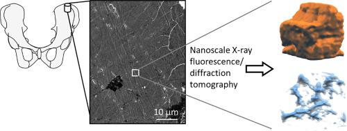
Studying nanostructured hierarchical materials such as the biomineralized bone is challenging due to their complex 3D structures that call for high spatial resolution. One route to study such materials is X-ray powder diffraction computed tomography (XRD-CT) that reveals the 3D distribution of crystalline phases and X-ray fluorescence computed tomography (XRF-CT) that provides element distributions. However, the spatial resolution of XRD-CT has thus far been limited. Here we demonstrate better than 120 nm 3D resolution on human bone in XRD-CT and XRF-CT measured simultaneously using X-ray nanobeams. The results pave the way for nanoscale 3D characterization of nanocrystalline composites like bone at unprecedented detail.
中文翻译:
对人体骨骼进行纳米束 X 射线荧光和衍射计算机断层扫描,分辨率优于 120 nm
由于其复杂的 3D 结构需要高空间分辨率,因此研究纳米结构分层材料(例如生物矿化骨)具有挑战性。研究此类材料的一种途径是 X 射线粉末衍射计算机断层扫描 (XRD-CT),它揭示了晶相的 3D 分布,以及提供元素分布的 X 射线荧光计算机断层扫描 (XRF-CT)。然而,迄今为止,XRD-CT 的空间分辨率受到限制。在这里,我们在使用 X 射线纳米束同时测量的 XRD-CT 和 XRF-CT 中证明了人体骨骼的 3D 分辨率优于 120 nm。结果为以前所未有的细节对骨骼等纳米晶复合材料进行纳米级 3D 表征铺平了道路。


 京公网安备 11010802027423号