iScience ( IF 4.6 ) Pub Date : 2020-09-19 , DOI: 10.1016/j.isci.2020.101590 Lorin Timaeus , Laura Geid , Gizem Sancer , Mathias F. Wernet , Thomas Hummel
|
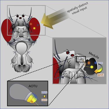
One hallmark of the visual system is a strict retinotopic organization from the periphery toward the central brain, where functional imaging in Drosophila revealed a spatially accurate representation of visual cues in the central complex. This raised the question how, on a circuit level, the topographic features are implemented, as the majority of visual neurons enter the central brain converge in optic glomeruli. We discovered a spatial segregation of topographic versus nontopographic projections of distinct classes of medullo-tubercular (MeTu) neurons into a specific visual glomerulus, the anterior optic tubercle (AOTU). These parallel channels synapse onto different tubercular-bulbar (TuBu) neurons, which in turn relay visual information onto specific central complex ring neurons in the bulb neuropil. Hence, our results provide the circuit basis for spatially accurate representation of visual information and highlight the AOTU's role as a prominent relay station for spatial information from the retina to the central brain.
中文翻译:
具有地形和非地形组织的并行视觉通路将果蝇眼连接到中央大脑
视觉系统的一个标志是从外围到中央大脑的严格视网膜组织,果蝇的功能成像揭示了中央建筑群中视觉线索的空间精确表示。这就提出了一个问题,即如何在电路水平上实现地形特征,因为大多数视觉神经元进入中枢神经会聚在视神经小球中。我们发现了不同类别的延髓-肾小管(MeTu)神经元的地形与非地形投影的空间分离,将其分成特定的视觉肾小球,前视神经结节(AOTU)。这些平行通道突触到不同的结核球(TuBu)神经元上,后者又将视觉信息传递到灯泡神经枕中的特定中央复杂环状神经元上。因此,我们的结果为视觉信息的空间精确表示提供了电路基础,并突出了AOTU'


 京公网安备 11010802027423号