Spectrochimica Acta Part A: Molecular and Biomolecular Spectroscopy ( IF 4.3 ) Pub Date : 2020-09-16 , DOI: 10.1016/j.saa.2020.118916 Caroline Moore , Andrew Harvey , Jonathan N. Coleman , Hugh J. Byrne , Jennifer McIntyre
|
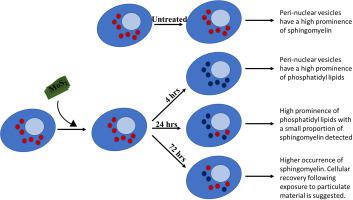
The emergence of large scale production techniques for 2D particulate materials has dramatically increased their applications potential. Understanding the interactions of biological cells with such particulate material is therefore of paramount importance, both for toxicological assessment and potential biomedical applications. Conventional in-vitro cytological assays commonly record only a single colorimetric end-point, and do not provide an in-depth analysis of how such materials are uptaken and processed within cells. To demonstrate its potential as an alternative, label free approach, confocal Raman micro-spectroscopy has been used to profile the cellular response of macrophage-like immune cells as a result of exposure to a sub-lethal dose of particulate MoS2, as an example novel 2D material. Particles were seen to be uptaken and trafficked in sub-cellular vesicles, and this sensitive technique allows differences in the biochemical composition of the vesicles to be assessed and monitored as a function of time. Untreated macrophage-like cells contain lipidic vesicles which are found to be relatively rich in the membrane lipid sphingomyelin, key to the process of cell membrane regeneration. After exposure to MoS2, the particulate material is seen to be invaginated in similar vesicles, the most prominent of which now, however, have spectroscopic signatures which are dominated by those of phosphatidyl family lipids, consistent with the phagocytotic pathway. The lipidic content of cells is seen to increase at all time-points (4, 24 and 72 h). although vesicles composed of sphingomyelin become more prominent again following a prolonged incubation of 72 h to a sub-lethal dose of MoS2, as the immune cell has processed the particulate material and initiates recovery to a normal/untreated state. This study reveals Raman micro-spectroscopy is an effective method for monitoring cellular responses and evolution of organelle compositions in response to MoS2 exposure. The additional benefit of using this technique is that cells can be monitored as a function of time, while it can also be used for screening other micro/nano materials for toxicology and/or establishing cell responses.
中文翻译:
MoS 2暴露后使用拉曼显微光谱技术无标记筛选巨噬细胞样细胞中的生化变化
二维颗粒材料的大规模生产技术的出现极大地增加了其应用潜力。因此,对于毒理学评估和潜在的生物医学应用而言,了解生物细胞与此类颗粒物质的相互作用至关重要。常规的体外细胞学测定通常仅记录单个比色终点,并且不提供对如何在细胞内摄取和加工此类物质的深入分析。为了证明其作为替代,无标记方法的潜力,共聚焦拉曼显微光谱法已被用于分析巨噬细胞样免疫细胞由于暴露于致死剂量的颗粒状MoS 2而产生的细胞反应。,例如新颖的2D材料。可见颗粒在亚细胞小泡中被摄取和运输,并且这种灵敏的技术允许根据时间评估和监测小泡的生化组成的差异。未经处理的巨噬细胞样细胞含有脂质小泡,发现脂质小泡中的膜脂质鞘磷脂相对丰富,这是细胞膜再生过程的关键。暴露于MoS 2后可见,颗粒物质被掺入到类似的囊泡中,然而,其中最突出的是具有光谱特征,该特征由磷脂酰家族脂质的特征所主导,与吞噬途径一致。可见细胞的脂质含量在所有时间点(4、24和72小时)均增加。尽管由鞘磷脂组成的囊泡在亚致死剂量的MoS 2长时间孵育72小时后再次变得更加突出,因为免疫细胞已经处理了颗粒物质并开始恢复至正常/未处理状态。这项研究表明拉曼显微光谱法是监测细胞响应和响应MoS 2的细胞器组成演变的有效方法接触。使用该技术的另一个好处是,可以根据时间监视细胞,同时还可以用于筛选其他微/纳米材料的毒理学和/或建立细胞反应。


 京公网安备 11010802027423号