当前位置:
X-MOL 学术
›
STEM CELLS
›
论文详情
Our official English website, www.x-mol.net, welcomes your feedback! (Note: you will need to create a separate account there.)
Activation of β-catenin by TGF-β1 promotes ligament-fibroblastic differentiation and inhibits cementoblastic differentiation of human periodontal ligament cells
STEM CELLS ( IF 5.2 ) Pub Date : 2020-09-15 , DOI: 10.1002/stem.3275 Jong-Chan Lim 1 , Sang-Hoon Bae 1 , Gyutae Lee 2 , Chun Jeih Ryu 3 , Young-Joo Jang 1, 4
STEM CELLS ( IF 5.2 ) Pub Date : 2020-09-15 , DOI: 10.1002/stem.3275 Jong-Chan Lim 1 , Sang-Hoon Bae 1 , Gyutae Lee 2 , Chun Jeih Ryu 3 , Young-Joo Jang 1, 4
Affiliation
|
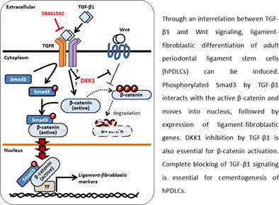
TGF‐β and Wnt/β‐catenin signaling pathways are known to be essential for the development of periodontal tissue. In this study, we examined the crosstalk between TGF‐β and Wnt/β‐catenin signaling in ligament‐fibroblastic differentiation of human periodontal ligament cells (hPDLCs). TGF‐β1 treatment significantly increased the expression of ligament‐fibroblastic markers, but such expression was preventing by treatment with SB431542, a TGF‐β type I receptor inhibitor. As well as phosphorylation of Smad3, TGF‐β1 increased β‐catenin activation. The depletion of β‐catenin reduced the expression of ligament‐fibroblastic markers, suggesting that β‐catenin is essential for ligament differentiation. The effect of TGF‐β1 on β‐catenin activation did not seem to be much correlated with Wnt stimuli, but endogenous DKK1 was suppressed by TGF‐β1, indicating that β‐catenin activation could be increased much more by TGF‐β1. In addition to DKK1 suppression, Smad3 phosphorylation by TGF‐β1 facilitated the nuclear translocation of cytoplasmic β‐catenin. In contrast to ligament‐fibroblastic differentiation, inhibition of TGF‐β1 signaling was needed for cementoblastic differentiation of hPDLCs. BMP7 treatment accompanied by inhibition of TGF‐β1 signaling had a synergistic effect on cementoblastic differentiation. In conclusion, β‐catenin activation by TGF‐β1 caused ligament‐fibroblastic differentiation of hPDLCs, and the presence of TGF‐β1 stimuli basically determined whether hPDLCs are differentiated into ligament progenitor or cementoblasts.
中文翻译:

TGF-β1激活β-catenin促进韧带-成纤维细胞分化并抑制人牙周膜细胞成牙骨质细胞分化
已知 TGF-β 和 Wnt/β-catenin 信号通路对牙周组织的发育至关重要。在这项研究中,我们检查了人牙周膜细胞 (hPDLCs) 的韧带成纤维细胞分化中 TGF-β 和 Wnt/β-catenin 信号之间的串扰。TGF-β1 治疗显着增加了韧带成纤维细胞标志物的表达,但这种表达被 SB431542(一种 TGF-β I 型受体抑制剂)治疗所阻止。除了 Smad3 的磷酸化外,TGF-β1 还增加了 β-catenin 的活化。β-catenin 的消耗降低了韧带成纤维细胞标志物的表达,表明 β-catenin 是韧带分化所必需的。TGF-β1 对 β-catenin 激活的影响似乎与 Wnt 刺激没有太大关系,但内源性 DKK1 被 TGF-β1 抑制,表明 TGF-β1 可以更多地增加 β-连环蛋白的活化。除了抑制 DKK1 外,TGF-β1 对 Smad3 的磷酸化促进了细胞质 β-连环蛋白的核易位。与韧带-成纤维细胞分化相反,hPDLCs 的成牙骨质细胞分化需要抑制 TGF-β1 信号。BMP7 处理伴随着 TGF-β1 信号的抑制对成牙骨质细胞分化具有协同作用。总之,TGF-β1 激活 β-catenin 导致 hPDLCs 的韧带-成纤维细胞分化,TGF-β1 刺激的存在基本上决定了 hPDLCs 是否分化为韧带祖细胞或成牙骨质细胞。TGF-β1 对 Smad3 的磷酸化促进了细胞质 β-连环蛋白的核易位。与韧带-成纤维细胞分化相反,hPDLCs 的成牙骨质细胞分化需要抑制 TGF-β1 信号。BMP7 处理伴随着 TGF-β1 信号的抑制对成牙骨质细胞分化具有协同作用。总之,TGF-β1 激活 β-catenin 导致 hPDLCs 的韧带-成纤维细胞分化,TGF-β1 刺激的存在基本上决定了 hPDLCs 是否分化为韧带祖细胞或成牙骨质细胞。TGF-β1 对 Smad3 的磷酸化促进了细胞质 β-catenin 的核转位。与韧带-成纤维细胞分化相反,hPDLCs 的成牙骨质细胞分化需要抑制 TGF-β1 信号。BMP7 处理伴随着 TGF-β1 信号的抑制对成牙骨质细胞分化具有协同作用。总之,TGF-β1 激活 β-catenin 导致 hPDLCs 的韧带-成纤维细胞分化,TGF-β1 刺激的存在基本上决定了 hPDLCs 是否分化为韧带祖细胞或成牙骨质细胞。BMP7 处理伴随着 TGF-β1 信号的抑制对成牙骨质细胞分化具有协同作用。总之,TGF-β1 激活 β-catenin 导致 hPDLCs 的韧带-成纤维细胞分化,TGF-β1 刺激的存在基本上决定了 hPDLCs 是否分化为韧带祖细胞或成牙骨质细胞。BMP7 处理伴随着 TGF-β1 信号的抑制对成牙骨质细胞分化具有协同作用。总之,TGF-β1 激活 β-catenin 导致 hPDLCs 的韧带-成纤维细胞分化,TGF-β1 刺激的存在基本上决定了 hPDLCs 是否分化为韧带祖细胞或成牙骨质细胞。
更新日期:2020-09-15
中文翻译:
TGF-β1激活β-catenin促进韧带-成纤维细胞分化并抑制人牙周膜细胞成牙骨质细胞分化
已知 TGF-β 和 Wnt/β-catenin 信号通路对牙周组织的发育至关重要。在这项研究中,我们检查了人牙周膜细胞 (hPDLCs) 的韧带成纤维细胞分化中 TGF-β 和 Wnt/β-catenin 信号之间的串扰。TGF-β1 治疗显着增加了韧带成纤维细胞标志物的表达,但这种表达被 SB431542(一种 TGF-β I 型受体抑制剂)治疗所阻止。除了 Smad3 的磷酸化外,TGF-β1 还增加了 β-catenin 的活化。β-catenin 的消耗降低了韧带成纤维细胞标志物的表达,表明 β-catenin 是韧带分化所必需的。TGF-β1 对 β-catenin 激活的影响似乎与 Wnt 刺激没有太大关系,但内源性 DKK1 被 TGF-β1 抑制,表明 TGF-β1 可以更多地增加 β-连环蛋白的活化。除了抑制 DKK1 外,TGF-β1 对 Smad3 的磷酸化促进了细胞质 β-连环蛋白的核易位。与韧带-成纤维细胞分化相反,hPDLCs 的成牙骨质细胞分化需要抑制 TGF-β1 信号。BMP7 处理伴随着 TGF-β1 信号的抑制对成牙骨质细胞分化具有协同作用。总之,TGF-β1 激活 β-catenin 导致 hPDLCs 的韧带-成纤维细胞分化,TGF-β1 刺激的存在基本上决定了 hPDLCs 是否分化为韧带祖细胞或成牙骨质细胞。TGF-β1 对 Smad3 的磷酸化促进了细胞质 β-连环蛋白的核易位。与韧带-成纤维细胞分化相反,hPDLCs 的成牙骨质细胞分化需要抑制 TGF-β1 信号。BMP7 处理伴随着 TGF-β1 信号的抑制对成牙骨质细胞分化具有协同作用。总之,TGF-β1 激活 β-catenin 导致 hPDLCs 的韧带-成纤维细胞分化,TGF-β1 刺激的存在基本上决定了 hPDLCs 是否分化为韧带祖细胞或成牙骨质细胞。TGF-β1 对 Smad3 的磷酸化促进了细胞质 β-catenin 的核转位。与韧带-成纤维细胞分化相反,hPDLCs 的成牙骨质细胞分化需要抑制 TGF-β1 信号。BMP7 处理伴随着 TGF-β1 信号的抑制对成牙骨质细胞分化具有协同作用。总之,TGF-β1 激活 β-catenin 导致 hPDLCs 的韧带-成纤维细胞分化,TGF-β1 刺激的存在基本上决定了 hPDLCs 是否分化为韧带祖细胞或成牙骨质细胞。BMP7 处理伴随着 TGF-β1 信号的抑制对成牙骨质细胞分化具有协同作用。总之,TGF-β1 激活 β-catenin 导致 hPDLCs 的韧带-成纤维细胞分化,TGF-β1 刺激的存在基本上决定了 hPDLCs 是否分化为韧带祖细胞或成牙骨质细胞。BMP7 处理伴随着 TGF-β1 信号的抑制对成牙骨质细胞分化具有协同作用。总之,TGF-β1 激活 β-catenin 导致 hPDLCs 的韧带-成纤维细胞分化,TGF-β1 刺激的存在基本上决定了 hPDLCs 是否分化为韧带祖细胞或成牙骨质细胞。


 京公网安备 11010802027423号