当前位置:
X-MOL 学术
›
Prog. Earth Planet. Sci.
›
论文详情
Our official English website, www.x-mol.net, welcomes your feedback! (Note: you will need to create a separate account there.)
Visualization of the morphology and mode of occurrence of Cenomanian rudists within a drillcore by X-ray CT scanning and 3D modeling
Progress in Earth and Planetary Science ( IF 3.9 ) Pub Date : 2020-09-14 , DOI: 10.1186/s40645-020-00359-7 Motoyoshi Yamanaka , Shin-ichi Sano , Hamad Bu Alrougha Al Zaabi , Hiroshi Fujioka , Yasufumi Iryu
Progress in Earth and Planetary Science ( IF 3.9 ) Pub Date : 2020-09-14 , DOI: 10.1186/s40645-020-00359-7 Motoyoshi Yamanaka , Shin-ichi Sano , Hamad Bu Alrougha Al Zaabi , Hiroshi Fujioka , Yasufumi Iryu
|
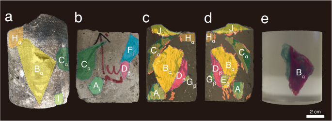
Rudists are a group of bizarrely shaped marine bivalves that lived in
the Tethys Ocean from the Late Jurassic to the latest Cretaceous. They are
morphologically variable, including snail-like, cup-like, and horn-like shapes. In
the Middle East, Cretaceous carbonates with rudists and rudist fragments are well
exposed in many outcrops as well as oil and gas fields. For rudist-bearing
carbonates in the subsurface, knowledge of rudist morphology and mode of occurrence
must be derived from observation of drillcores; however, understanding the
three-dimensional (3D) geometry of rudists from observation of core surfaces is
difficult. In paleontological studies of rudists, X-ray computerized tomography (CT)
scans have been carried out to reconstruct the inside texture of rudist shells for
the purpose of taxonomic research. In contrast, in the oil and gas industry,
application of X-ray CT scanning technology is generally focused on direct
measurement of reservoir properties. Studies of rudist fossils within drillcores by
means of X-ray CT have not yet been conducted. We have developed a new protocol to
observe core interiors using X-ray CT. We obtained high-resolution 3D images of
rudists in a drillcore by means of surface rendering, volume rendering, and 3D
printing. X-ray CT and 3D modeling is a novel method for non-destructive analyses of
the morphology and mode of occurrence of fossils within drillcores.

中文翻译:

通过X射线CT扫描和3D建模可视化钻芯内的Cenomanian ruders的形态和发生方式
鲁迪特人是一群怪异的海洋双壳类动物,它们生活在侏罗纪晚期至最新的白垩纪的特提斯海洋中。它们的形态是可变的,包括蜗牛形,杯形和角形。在中东,在许多露头以及油气田中,暴露有白垩纪碳酸盐岩并伴有鲁氏岩和鲁氏岩碎片。对于地下带轴承的碳酸盐岩,必须从对钻芯的观察中得出有关其形态和发生方式的知识。但是,很难通过观察核心表面来了解三叉戟的三维(3D)几何形状。在红树林的古生物学研究中,为了进行分类学研究,已经进行了X射线计算机断层扫描(CT)扫描以重建红树林的内部纹理。相反,在石油和天然气工业中,X射线CT扫描技术的应用通常侧重于直接测量储层物性。尚未进行通过X射线CT进行钻芯内的红土化石研究。我们已经开发出一种新的协议,可以使用X射线CT观察核心内部。通过表面渲染,体绘制和3D打印,我们在钻芯中获得了高分辨率的3D立体图。X射线CT和3D建模是一种新颖的方法,可对钻芯内化石的形态和发生模式进行无损分析。我们已经开发出一种新的协议,可以使用X射线CT观察核心内部。通过表面渲染,体绘制和3D打印,我们在钻芯中获得了高分辨率的3D立体图。X射线CT和3D建模是一种新颖的方法,可对钻芯内化石的形态和发生模式进行无损分析。我们已经开发出一种新的协议,可以使用X射线CT观察核心内部。通过表面渲染,体绘制和3D打印,我们在钻芯中获得了高分辨率的3D立体图。X射线CT和3D建模是一种新颖的方法,可对钻芯内化石的形态和发生模式进行无损分析。
更新日期:2020-09-14

中文翻译:
通过X射线CT扫描和3D建模可视化钻芯内的Cenomanian ruders的形态和发生方式
鲁迪特人是一群怪异的海洋双壳类动物,它们生活在侏罗纪晚期至最新的白垩纪的特提斯海洋中。它们的形态是可变的,包括蜗牛形,杯形和角形。在中东,在许多露头以及油气田中,暴露有白垩纪碳酸盐岩并伴有鲁氏岩和鲁氏岩碎片。对于地下带轴承的碳酸盐岩,必须从对钻芯的观察中得出有关其形态和发生方式的知识。但是,很难通过观察核心表面来了解三叉戟的三维(3D)几何形状。在红树林的古生物学研究中,为了进行分类学研究,已经进行了X射线计算机断层扫描(CT)扫描以重建红树林的内部纹理。相反,在石油和天然气工业中,X射线CT扫描技术的应用通常侧重于直接测量储层物性。尚未进行通过X射线CT进行钻芯内的红土化石研究。我们已经开发出一种新的协议,可以使用X射线CT观察核心内部。通过表面渲染,体绘制和3D打印,我们在钻芯中获得了高分辨率的3D立体图。X射线CT和3D建模是一种新颖的方法,可对钻芯内化石的形态和发生模式进行无损分析。我们已经开发出一种新的协议,可以使用X射线CT观察核心内部。通过表面渲染,体绘制和3D打印,我们在钻芯中获得了高分辨率的3D立体图。X射线CT和3D建模是一种新颖的方法,可对钻芯内化石的形态和发生模式进行无损分析。我们已经开发出一种新的协议,可以使用X射线CT观察核心内部。通过表面渲染,体绘制和3D打印,我们在钻芯中获得了高分辨率的3D立体图。X射线CT和3D建模是一种新颖的方法,可对钻芯内化石的形态和发生模式进行无损分析。


 京公网安备 11010802027423号