Journal of Neuroscience Methods ( IF 2.7 ) Pub Date : 2020-09-12 , DOI: 10.1016/j.jneumeth.2020.108946 Sibel Çimen Yetiş 1 , Abdulkerim Çapar 2 , Dursun A Ekinci 2 , Umut E Ayten 1 , Bilal E Kerman 3 , B Uğur Töreyin 2
|
Background
The myelin sheath produced by glial cells insulates the axons, and supports the function of the nervous system. Myelin sheath degeneration causes neurodegenerative disorders, such as multiple sclerosis (MS). There are no therapies for MS that promote remyelination. Drug discovery frequently involves screening thousands of compounds. However, this is not feasible for remyelination drugs, since myelin quantification is a manual labor-intensive endeavor. Therefore, the development of assistive software for expedited myelin detection is instrumental for MS drug discovery by enabling high-content image-based drug screens.
New method
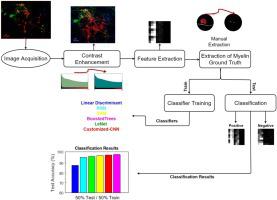
In this study, we developed a machine learning based expedited myelin detection approach in fluorescence microscopy images. Multi-channel three-dimensional microscopy images of a mouse stem cell-based myelination assay were labeled by experts. A spectro-spatial feature extraction method was introduced to represent local dependencies of voxels both in spatial and spectral domains. Feature extraction yielded two data set of over forty-seven thousand annotated images in total.
Results
Myelin detection performances of 23 different supervised machine learning techniques including a customized-convolutional neural network (CNN), were assessed using various train/test split ratios of the data sets. The highest accuracy values of and were achieved by Boosted Trees and customized-CNN, respectively.
Comparison with existing methods
Our approach can detect myelin in a common experimental setup. Myelin extending in any orientation in 3 dimensions is segmented from 3 channel z-stack fluorescence images.
Conclusions
Our results suggest that the proposed expedited myelin detection approach is a feasible and robust method for remyelination drug screening.
中文翻译:
使用机器学习检测荧光显微镜图像中的髓磷脂。
背景
由神经胶质细胞产生的髓鞘可隔离轴突,并支持神经系统的功能。髓鞘变性引起神经退行性疾病,例如多发性硬化症(MS)。没有用于促进髓鞘再生的MS疗法。药物发现经常涉及筛选数千种化合物。但是,这对于髓鞘再生药物是不可行的,因为髓磷脂的定量是一项人工劳动密集型的工作。因此,通过实现高含量的基于图像的药物筛选,用于加速髓磷脂检测的辅助软件的开发对于MS药物发现至关重要。
新方法
在这项研究中,我们开发了一种基于机器学习的荧光显微镜图像中快速的髓磷脂检测方法。专家标记了基于小鼠干细胞的髓鞘形成测定的多通道三维显微图像。引入了光谱空间特征提取方法来表示体素在空间和光谱域中的局部依赖性。特征提取产生了两个数据集,总共超过47千个带注释的图像。
结果
使用数据集的各种训练/测试拆分率评估了23种不同的监督式机器学习技术(包括定制卷积神经网络(CNN))的髓磷脂检测性能。的最高精度值 和 分别由Boosted Trees和定制的CNN实现。
与现有方法的比较
我们的方法可以在常见的实验装置中检测髓磷脂。从3通道z-stack荧光图像中分割了在3个维度上以任何方向延伸的髓磷脂。
结论
我们的结果表明,提出的快速髓磷脂检测方法是一种可行且健壮的再髓鞘药物筛选方法。


 京公网安备 11010802027423号