Acta Biomaterialia ( IF 9.4 ) Pub Date : 2020-09-12 , DOI: 10.1016/j.actbio.2020.09.004 Jean-Baptiste Forien 1 , Jun Uzuhashi 2 , Tadakatsu Ohkubo 2 , Kazuhiro Hono 2 , Lucy Luo 3 , Henry P Schwarcz 4 , Alix C Deymier 5 , Christina Krywka 6 , Claudia Fleck 7 , Paul Zaslansky 8
|
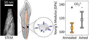
Bone-like materials comprise carbonated-hydroxyapatite nanocrystals (c-Ap) embedding a fibrillar collagen matrix. The mineral particles stiffen the nanocomposite by tight attachment to the protein fibrils creating a high strength and toughness material. The nanometer dimensions of c-Ap crystals make it very challenging to measure their mechanical properties. Mineral in bony tissues such as dentine contains 2~6 wt.% carbonate with possibly different elastic properties as compared with crystalline hydroxyapatite. Here we determine strain in biogenic apatite nanocrystals by directly measuring atomic deformation in pig dentine before and after removing carbonate. Transmission electron microscopy revealed the platy 3D morphology while atom probe tomography revealed carbon inside the calcium rich domains. High-energy X-ray diffraction in combination with in situ hydrostatic pressurization quantified reversible c-Ap deformations. Crystal strains differed between annealed and ashed (decarbonated) samples, following 1 or 10 h heating at 250 °C or 550 °C respectively. Measured bulk moduli (K) and a-/c-lattice deformation ratios (η) were used to generate synthetic Ksyn and ηsyn identifying the most likely elastic constants C33 and C13 for c-Ap. These were then used to calculate the nanoparticle elastic moduli. For ashed samples, we find an average E11=107 GPa and E33 =128 GPa corresponding to ~5% and ~17% stiffening of the a-/c-axes of the nanocrystals as compared with the biogenic nanocrystals in annealed samples. Ashed samples exhibit ~10% lower Poisson's ratios as compared with the 0.25~0.36 range of carbonated apatite. Carbonate in c-Ap may therefore serve for tuning local deformability within bony tissues.
Statement of Significance
Carbonated apatite nanoparticles, typical for bony tissues, stiffen the network of collagen fibrils. However, it is not known if the biogenic apatite mechanical (elastic) properties differ from those of geologic mineral counterparts. Indeed the tiny dimensions and variable carbonate composition may have strong effects on deformation resistance. The present study provides experimental measurements of the elastic constants which we use to estimate Young's moduli and Poisson's ratio values. Comparison between ashed and annealed dentine samples quantifies the properties of both carbonated and decarbonated apatite nanocrystals. The results reveal fundamental attributes of bony mineral and showcase the additive advantages of combining X-ray diffraction with in situ hydrostatic compression, backed by atom probe and transmission electron microscopy tomography.
中文翻译:
牙本质磷灰石纳米晶体的X射线衍射和原位加压量化了去除碳酸盐后的模量刚度。
骨样材料包括嵌入纤维状胶原基质的碳酸羟基磷灰石纳米晶体(c-Ap)。矿物颗粒通过紧密附着在蛋白质原纤维上而使纳米复合材料变硬,从而形成高强度和韧性的材料。c-Ap晶体的纳米尺寸使测量其机械性能变得非常困难。与晶状羟基磷灰石相比,骨组织中的矿物质(例如牙本质)含有2〜6 wt。%的碳酸盐,其弹性可能不同。在这里,我们通过直接测量去除碳酸盐前后的猪牙本质中的原子变形来确定生物磷灰石纳米晶体中的应变。透射电子显微镜显示出板状3D形态,而原子探针层析成像显示出富含钙的区域内的碳。原位静水加压量化了可逆的c-Ap变形。分别在250°C或550°C加热1或10 h后,退火和灰化(脱碳)样品的晶体应变有所不同。测量体积弹性模量(ķ)和A- / C-晶格变形(比率η)被用来生成合成ķ顺式和η顺识别最有可能的弹性常数Ç 33和Ç 13对c-鸭。然后将这些用于计算纳米粒子的弹性模量。对于灰烬样品,我们发现平均E 11= 107 GPa和E 33 = 128 GPa,与退火样品中的生物纳米晶体相比,纳米晶体a- / c-轴的〜5%和〜17%硬化。与灰化磷灰石的0.25〜0.36范围相比,灰化的样品的泊松比降低了约10%。因此,c-Ap中的碳酸盐可用于调整骨组织内的局部变形能力。
重要声明
碳化磷灰石纳米颗粒(通常用于骨骼组织)会增强胶原纤维的网络。但是,尚不知道生物磷灰石的机械(弹性)特性是否与地质矿物对应的特性不同。确实,微小的尺寸和可变的碳酸盐成分可能会对抗变形性产生强烈影响。本研究提供了弹性常数的实验测量值,我们用它们来估计杨氏模量和泊松比值。牙本质样品和退火牙本质样品之间的比较可量化碳酸盐和脱碳磷灰石纳米晶体的性能。结果揭示了骨矿物的基本属性,并展示了将X射线衍射与原位相结合的附加优势 静水压缩,以原子探针和透射电子显微镜断层扫描为后盾。


 京公网安备 11010802027423号