Our official English website, www.x-mol.net, welcomes your
feedback! (Note: you will need to create a separate account there.)
Janus nanoparticles designed for extended cell surface attachment.
Nanoscale ( IF 5.8 ) Pub Date : 2020-09-11 , DOI: 10.1039/d0nr04061d Reshma Kadam 1 , Jaee Ghawali , Mario Waespy , Michael Maas , Kurosch Rezwan
Nanoscale ( IF 5.8 ) Pub Date : 2020-09-11 , DOI: 10.1039/d0nr04061d Reshma Kadam 1 , Jaee Ghawali , Mario Waespy , Michael Maas , Kurosch Rezwan
Affiliation
|
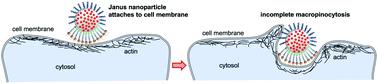
In this study, we present Janus nanoparticles that are designed for attaching to a eukaryotic cell surface with minimal cell uptake. This contrasts the rapid uptake via various endocytosis pathways that non-passivated isotropic particles usually encounter. Firmly attaching nanoparticles onto cell surfaces for extended periods of time can be a powerful new strategy to employ functional properties of nanoparticles for non-invasive interrogation and manipulation of biological systems. To this end, we synthesized rhodamine-doped silica (SiO2) nanoparticles functionalized with 1,2-distearoyl-sn-glycero-3-phosphoethanolamine (DSPE) on one hemisphere of the nanoparticle surface and high-molecular-weight long-chain poly(ethylene glycol) on the other one using the wax-Pickering emulsion technique. Nanoparticle localization was studied with NIH 3T3 rat fibroblasts in vitro. In these studies, the Janus nanoparticles adhered to the cell surface and, in contrast to isotropic control particles, only negligible uptake into the cells was observed, even after 24 h of incubation. In order to characterize the potential endocytosis pathway involved in the uptake of the Janus nanoparticles in more detail, fibroblasts and nanoparticles were incubated in the presence or absence of different endocytosis inhibitors. Our findings indicate that the Janus particles are not affected by caveolae- and receptor-mediated endocytosis and the prolonged attachment of the Janus nanoparticles is most likely the result of an incomplete macropinocytosis process. Consequently, by design, these Janus nanoparticles have the potential to firmly anchor onto cell surfaces for extended periods of time and might be utilized in various biotechnological and biomedical applications like cell surface tagging, magnetic manipulation of the cell membrane or non-invasive drug and gene delivery.
中文翻译:

Janus纳米粒子设计用于延长细胞表面附着。
在这项研究中,我们介绍了Janus纳米粒子,该纳米粒子设计用于以最小的细胞摄取附着到真核细胞表面。这与非钝化各向同性颗粒通常通过各种内吞途径快速吸收形成鲜明对比。长时间将纳米颗粒牢固地附着在细胞表面可能是一种强大的新策略,可以利用纳米颗粒的功能特性进行生物系统的非侵入式询问和操作。为此,我们合成了用1,2-二硬脂酰-sn-甘油官能化的若丹明掺杂的二氧化硅(SiO 2)纳米颗粒使用蜡提取乳液技术,在纳米颗粒表面的一个半球上放置-3-磷酸乙醇胺(DSPE),在另一个表面上放置一个高分子量长链聚乙二醇。使用NIH 3T3大鼠成纤维细胞体外研究了纳米粒子的定位。在这些研究中,Janus纳米颗粒粘附在细胞表面,与各向同性对照颗粒相反,即使在孵育24小时后,也只能观察到微不足道的细胞摄取。为了更详细地描述参与Janus纳米颗粒摄取的潜在内吞途径,在存在或不存在不同内吞抑制剂的情况下孵育成纤维细胞和纳米颗粒。我们的发现表明,Janus颗粒不受小窝和受体介导的内吞作用的影响,Janus纳米颗粒的长时间附着很可能是不完整的巨胞饮作用过程的结果。因此,通过设计,
更新日期:2020-09-24
中文翻译:
Janus纳米粒子设计用于延长细胞表面附着。
在这项研究中,我们介绍了Janus纳米粒子,该纳米粒子设计用于以最小的细胞摄取附着到真核细胞表面。这与非钝化各向同性颗粒通常通过各种内吞途径快速吸收形成鲜明对比。长时间将纳米颗粒牢固地附着在细胞表面可能是一种强大的新策略,可以利用纳米颗粒的功能特性进行生物系统的非侵入式询问和操作。为此,我们合成了用1,2-二硬脂酰-sn-甘油官能化的若丹明掺杂的二氧化硅(SiO 2)纳米颗粒使用蜡提取乳液技术,在纳米颗粒表面的一个半球上放置-3-磷酸乙醇胺(DSPE),在另一个表面上放置一个高分子量长链聚乙二醇。使用NIH 3T3大鼠成纤维细胞体外研究了纳米粒子的定位。在这些研究中,Janus纳米颗粒粘附在细胞表面,与各向同性对照颗粒相反,即使在孵育24小时后,也只能观察到微不足道的细胞摄取。为了更详细地描述参与Janus纳米颗粒摄取的潜在内吞途径,在存在或不存在不同内吞抑制剂的情况下孵育成纤维细胞和纳米颗粒。我们的发现表明,Janus颗粒不受小窝和受体介导的内吞作用的影响,Janus纳米颗粒的长时间附着很可能是不完整的巨胞饮作用过程的结果。因此,通过设计,


 京公网安备 11010802027423号