Journal of Structural Biology ( IF 3 ) Pub Date : 2020-09-11 , DOI: 10.1016/j.jsb.2020.107615 Dakota M Binkley 1 , Joseph Deering 2 , Hui Yuan 3 , Aurélien Gourrier 4 , Kathryn Grandfield 5
|
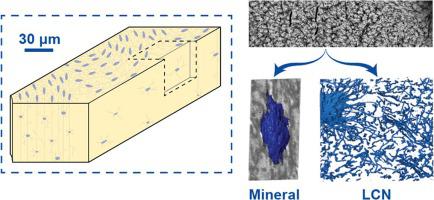
Visualizing bone mineralization and collagen fibril organization at intermediate scales between the nanometer and the hundreds of microns range, is still an important challenge. Similarly, visualizing cellular components which locally affect the tissue structure requires a precision of a few tens of nanometers at maximum while spanning several tens of micrometers. In the last decade, gallium focused ion beam (FIB) equipped with a scanning electron microscope (SEM) proved to be an extremely valuable structural tool to meet those ends. In this study, we assess the capability of a recent plasma FIB-SEM technology which provides a potential increase in measurement speed over gallium FIB-SEM, thus paving the way to larger volume analysis. Nanometer-scale layers of demineralized and mineralized unstained human femoral lamellar bone were sequentially sectioned over volumes of 6–16,000 μm3. Analysis of mineralized tissue revealed prolate ellipsoidal mineral clusters measuring approximately 1.1 µm in length by 700 nm at their maximum diameter. Those features, suggested by others in high resolution studies, appear here as a ubiquitous motif in mineralized lamellar bone over thousands of microns cubed, suggesting a heterogeneous and yet regular pattern of mineral deposition past the single collagen fibril level. This large scale view retained sufficient resolution to visualize the collagen fibrils while also partly visualizing the lacuno-canalicular network in three-dimensions. These findings are strong evidence for suitability of PFIB as a bone analysis tool and the need to revisit bone mineralization over multi-length scales with mineralized tissue.
中文翻译:
通过等离子体聚焦离子束连续切片在 3D 中显示人皮质骨中的椭圆体中尺度矿化模式。
在纳米和数百微米范围之间的中间尺度上可视化骨矿化和胶原纤维组织仍然是一个重要的挑战。类似地,局部影响组织结构的细胞成分的可视化需要最大几十纳米的精度,同时跨越几十微米。在过去十年中,配备扫描电子显微镜 (SEM) 的镓聚焦离子束 (FIB) 被证明是满足这些目标的极其有价值的结构工具。在这项研究中,我们评估了最近的等离子体 FIB-SEM 技术的能力,该技术提供了比镓 FIB-SEM 测量速度的潜在提高,从而为更大体积的分析铺平了道路。3 . 对矿化组织的分析显示,长椭圆形矿物簇长约 1.1 µm,最大直径为 700 nm。其他人在高分辨率研究中提出的这些特征,在这里作为一个无处不在的主题出现在超过数千微米立方的矿化层状骨中,表明矿物质沉积的异质性但规则模式超过单个胶原纤维水平。这种大尺度视图保留了足够的分辨率来可视化胶原纤维,同时也部分地在三个维度上可视化腔隙-小管网络。这些发现有力地证明了 PFIB 作为骨骼分析工具的适用性,以及重新审视具有矿化组织的多长度尺度上的骨骼矿化的必要性。


 京公网安备 11010802027423号