Developmental Cell ( IF 10.7 ) Pub Date : 2020-09-11 , DOI: 10.1016/j.devcel.2020.08.007 Fengzhu Xiong 1 , Wenzhe Ma 2 , Bertrand Bénazéraf 3 , L Mahadevan 4 , Olivier Pourquié 5
|
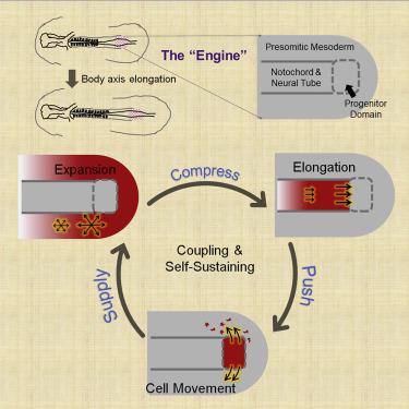
Tissues undergoing morphogenesis impose mechanical effects on one another. How developmental programs adapt to or take advantage of these effects remains poorly explored. Here, using a combination of live imaging, modeling, and microsurgical perturbations, we show that the axial and paraxial tissues in the forming avian embryonic body coordinate their rates of elongation through mechanical interactions. First, a cell motility gradient drives paraxial presomitic mesoderm (PSM) expansion, resulting in compression of the axial neural tube and notochord; second, elongation of axial tissues driven by PSM compression and polarized cell intercalation pushes the caudal progenitor domain posteriorly; finally, the axial push drives the lateral movement of midline PSM cells to maintain PSM growth and cell motility. These interactions form an engine-like positive feedback loop, which sustains a shared elongation rate for coupled tissues. Our results demonstrate a key role of inter-tissue forces in coordinating distinct body axis tissues during their co-elongation.
中文翻译:
机械耦合协调鸟类胚胎中轴向和旁轴组织的共同伸长。
经历形态发生的组织对彼此施加机械效应。发展计划如何适应或利用这些影响仍然缺乏探索。在这里,使用实时成像、建模和显微外科扰动的组合,我们展示了正在形成的禽类胚胎体中的轴向和旁轴组织通过机械相互作用协调它们的伸长率。首先,细胞运动梯度驱动旁轴前体中胚层 (PSM) 扩张,导致中轴神经管和脊索受压;其次,由 PSM 压缩和极化细胞插入驱动的轴向组织的伸长将尾部祖细胞域向后推动;最后,轴向推动驱动中线 PSM 细胞的横向运动,以维持 PSM 生长和细胞运动。这些相互作用形成了一个类似引擎的正反馈回路,维持了耦合组织的共享伸长率。我们的结果证明了组织间力在协调不同体轴组织的共同伸长过程中的关键作用。


 京公网安备 11010802027423号