Medical & Biological Engineering & Computing ( IF 2.6 ) Pub Date : 2020-08-25 , DOI: 10.1007/s11517-020-02216-7 Xiang Ying 1, 2 , Yulin Zhang 1, 2 , Mei Yu 1, 2 , Xi Wei 3 , Jialin Zhu 3 , Jie Gao 1, 2 , Zhiqiang Liu 1, 2 , Hongqian Shen 1, 2 , Ruixuan Zhang 1, 2 , Xuewei Li 1, 2 , Ruiguo Yu 1, 2
|
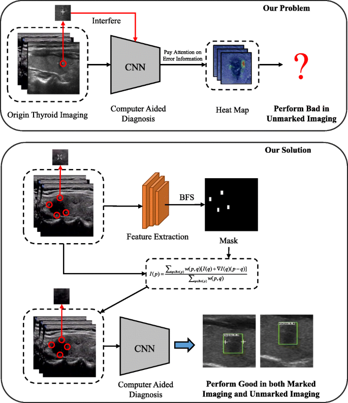
During thyroid ultrasound diagnosis, radiologists add markers such as pluses or crosses near a nodule’s edge to indicate the location of a nodule. For computer-aided detection, deep learning models achieve classification, segmentation, and detection by learning the thyroid’s texture in ultrasound images. Experiments show that manual markers are strong prior knowledge for data-driven deep learning models, which interferes with the judgment mechanism of computer-aided detection systems. Aiming at this problem, this paper proposes cascade marker removal algorithm for thyroid ultrasound images to eliminate the interference of manual markers. The algorithm consists of three parts. First, in order to highlight marked features, the algorithm extracts salient features in thyroid ultrasound images through feature extraction module. Secondly, mask correction module eliminates the interference of other features besides markers’ features. Finally, the marker removal module removes markers without destroying the semantic information in thyroid ultrasound images. Experiments show that our algorithm enables classification, segmentation, and object detection models to focus on the learning of pathological tissue features. At the same time, compared with mainstream image inpainting algorithms, our algorithm shows better performance on thyroid ultrasound images. In summary, our algorithm is of great significance for improving the stability and performance of computer-aided detection systems.
中文翻译:
甲状腺超声图像的级联标记去除算法。
在甲状腺超声诊断过程中,放射科医师会在结节边缘附近添加标记(例如加号或十字)以指示结节的位置。对于计算机辅助检测,深度学习模型通过学习超声图像中甲状腺的纹理来实现分类、分割和检测。实验表明,手动标记是数据驱动的深度学习模型的强先验知识,会干扰计算机辅助检测系统的判断机制。针对这一问题,本文提出了甲状腺超声图像级联标记去除算法,以消除人工标记的干扰。该算法由三部分组成。首先,为了突出标记特征,算法通过特征提取模块提取甲状腺超声图像中的显着特征。第二,掩模校正模块消除了标记特征之外的其他特征的干扰。最后,标记去除模块在不破坏甲状腺超声图像中的语义信息的情况下去除标记。实验表明,我们的算法使分类、分割和对象检测模型能够专注于病理组织特征的学习。同时,与主流图像修复算法相比,我们的算法在甲状腺超声图像上表现出更好的性能。总之,我们的算法对于提高计算机辅助检测系统的稳定性和性能具有重要意义。实验表明,我们的算法使分类、分割和对象检测模型能够专注于病理组织特征的学习。同时,与主流图像修复算法相比,我们的算法在甲状腺超声图像上表现出更好的性能。总之,我们的算法对于提高计算机辅助检测系统的稳定性和性能具有重要意义。实验表明,我们的算法使分类、分割和对象检测模型能够专注于病理组织特征的学习。同时,与主流图像修复算法相比,我们的算法在甲状腺超声图像上表现出更好的性能。总之,我们的算法对于提高计算机辅助检测系统的稳定性和性能具有重要意义。


 京公网安备 11010802027423号