Our official English website, www.x-mol.net, welcomes your
feedback! (Note: you will need to create a separate account there.)
Biomolecular Ultrasound Imaging of Phagolysosomal Function.
ACS Nano ( IF 15.8 ) Pub Date : 2020-09-09 , DOI: 10.1021/acsnano.0c05912 Bill Ling 1 , Justin Lee 2 , David Maresca 1 , Audrey Lee-Gosselin 1 , Dina Malounda 1 , Margaret B Swift 1 , Mikhail G Shapiro 1
ACS Nano ( IF 15.8 ) Pub Date : 2020-09-09 , DOI: 10.1021/acsnano.0c05912 Bill Ling 1 , Justin Lee 2 , David Maresca 1 , Audrey Lee-Gosselin 1 , Dina Malounda 1 , Margaret B Swift 1 , Mikhail G Shapiro 1
Affiliation
|
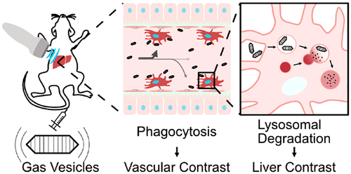
Phagocytic clearance and lysosomal processing of pathogens and debris are essential functions of the innate immune system. However, the assessment of these functions in vivo is challenging because most nanoscale contrast agents compatible with noninvasive imaging techniques are made from nonbiodegradable synthetic materials that do not undergo regular lysosomal degradation. To overcome this challenge, we describe the use of an all-protein contrast agent to directly visualize and quantify phagocytic and lysosomal activities in vivo by ultrasound imaging. This contrast agent is based on gas vesicles (GVs), a class of air-filled protein nanostructures naturally expressed by buoyant microbes. Using a combination of ultrasound imaging, pharmacology, immunohistology, and live-cell optical microscopy, we show that after intravenous injection, GVs are cleared from circulation by liver-resident macrophages. Once internalized, the GVs undergo lysosomal degradation, resulting in the elimination of their ultrasound contrast. By noninvasively monitoring the temporal dynamics of GV-generated ultrasound signal in circulation and in the liver and fitting them with a pharmacokinetic model, we can quantify the rates of phagocytosis and lysosomal degradation in living animals. We demonstrate the utility of this method by showing how these rates are perturbed in two models of liver dysfunction: phagocyte deficiency and nonalcoholic fatty liver disease. The combination of proteolytically degradable nanoscale contrast agents and quantitative ultrasound imaging thus enables noninvasive functional imaging of cellular degradative processes.
中文翻译:

吞噬溶酶体功能的生物分子超声成像。
病原体和碎片的吞噬清除和溶酶体处理是先天免疫系统的基本功能。然而,体内这些功能的评估具有挑战性,因为大多数与非侵入性成像技术兼容的纳米级造影剂是由不可生物降解的合成材料制成的,这些材料不会经历定期的溶酶体降解。为了克服这一挑战,我们描述了使用全蛋白造影剂通过超声成像直接可视化和量化体内吞噬细胞和溶酶体活动。这种造影剂基于气体囊泡 (GV),这是一类由浮力微生物自然表达的充满空气的蛋白质纳米结构。结合超声成像、药理学、免疫组织学和活细胞光学显微镜,我们发现静脉注射后,GV 被肝脏巨噬细胞从循环中清除。一旦内化,GV 就会经历溶酶体降解,导致其超声对比度消失。通过无创监测循环和肝脏中 GV 产生的超声信号的时间动态,并将其与药代动力学模型进行拟合,我们可以量化活体动物的吞噬作用和溶酶体降解速率。我们通过展示这些速率在两种肝功能障碍模型(吞噬细胞缺乏症和非酒精性脂肪肝病)中如何受到干扰来证明该方法的实用性。因此,可蛋白水解降解的纳米级造影剂和定量超声成像的结合使得能够对细胞降解过程进行无创功能成像。
更新日期:2020-09-22
中文翻译:
吞噬溶酶体功能的生物分子超声成像。
病原体和碎片的吞噬清除和溶酶体处理是先天免疫系统的基本功能。然而,体内这些功能的评估具有挑战性,因为大多数与非侵入性成像技术兼容的纳米级造影剂是由不可生物降解的合成材料制成的,这些材料不会经历定期的溶酶体降解。为了克服这一挑战,我们描述了使用全蛋白造影剂通过超声成像直接可视化和量化体内吞噬细胞和溶酶体活动。这种造影剂基于气体囊泡 (GV),这是一类由浮力微生物自然表达的充满空气的蛋白质纳米结构。结合超声成像、药理学、免疫组织学和活细胞光学显微镜,我们发现静脉注射后,GV 被肝脏巨噬细胞从循环中清除。一旦内化,GV 就会经历溶酶体降解,导致其超声对比度消失。通过无创监测循环和肝脏中 GV 产生的超声信号的时间动态,并将其与药代动力学模型进行拟合,我们可以量化活体动物的吞噬作用和溶酶体降解速率。我们通过展示这些速率在两种肝功能障碍模型(吞噬细胞缺乏症和非酒精性脂肪肝病)中如何受到干扰来证明该方法的实用性。因此,可蛋白水解降解的纳米级造影剂和定量超声成像的结合使得能够对细胞降解过程进行无创功能成像。


 京公网安备 11010802027423号