当前位置:
X-MOL 学术
›
NeuroImage
›
论文详情
Our official English website, www.x-mol.net, welcomes your feedback! (Note: you will need to create a separate account there.)
The human corticocortical vestibular network
NeuroImage ( IF 5.7 ) Pub Date : 2020-12-01 , DOI: 10.1016/j.neuroimage.2020.117362 T M Raiser 1 , V L Flanagin 1 , M Duering 2 , A van Ombergen 3 , R M Ruehl 4 , P Zu Eulenburg 5
NeuroImage ( IF 5.7 ) Pub Date : 2020-12-01 , DOI: 10.1016/j.neuroimage.2020.117362 T M Raiser 1 , V L Flanagin 1 , M Duering 2 , A van Ombergen 3 , R M Ruehl 4 , P Zu Eulenburg 5
Affiliation
|
BACKGROUND
Little is known about the cortical organization of human vestibular information processing. Instead of a dedicated primary vestibular cortex, a distributed network of regions across the cortex respond to vestibular input. The aim of this study is to characterize the human corticocortical vestibular network and compare it to established results in non-human primates. METHODS
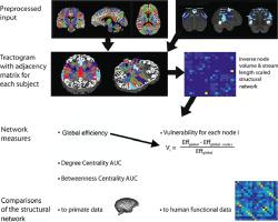
We collected high-resolution multi-shell diffusion-weighted (DWI) and state-of-the-art resting-state functional MR images of 29 right-handed normal subjects. Ten cortical vestibular regions per hemisphere were predefined from previous vestibular stimulation studies and applied as regions of interest. Four different structural corticocortical vestibular networks accounting for relevant constraints were investigated. The analyses included the investigation of common network measures and hemispheric differences for functional and structural connectivity patterns alike. In addition, the results of the structural vestibular network were compared to findings previously reported in non-human primates with respect to tracer injections (Guldin and Grusser, 1998). RESULTS
All structural networks independent of the applied constraints showed a recurring subdivision into identical three submodules. The structural human network was characterized by a predominantly intrahemispheric connectivity, whereas the functional pattern highlighted a strong connectivity for all homotopic nodes. A significant laterality preference towards the right hemisphere can be observed throughout the analyses: (1) with larger nodes, (2) stronger connectivity values structurally and functionally, and (3) a higher functional relevance. Similar connectivity patterns to non-human primate data were found in sensory and higher association cortices rather than premotor and motor areas. CONCLUSION
Our analysis delineated a remarkably stable organization of cortical vestibular connectivity. Differences found between primate species may be attributed to phylogeny as well as methodological differences. With our work we solidified evidence for lateralization within the corticocortical vestibular network. Our results might explain why cortical lesions in humans do not lead to persistent vestibular symptoms. Redundant structural routing throughout the network and a high-degree functional connectivity may buffer the network and reestablish network integrity quickly in case of injury.
中文翻译:

人类皮质前庭网络
背景对人类前庭信息处理的皮层组织知之甚少。不是专用的初级前庭皮层,而是分布在整个皮层的区域网络对前庭输入做出反应。本研究的目的是表征人类皮质前庭网络,并将其与非人类灵长类动物的既定结果进行比较。方法我们收集了 29 名惯用右手的正常受试者的高分辨率多壳扩散加权 (DWI) 和最先进的静息状态功能 MR 图像。每个半球的 10 个皮质前庭区域是从先前的前庭刺激研究中预先定义的,并作为感兴趣的区域应用。研究了四种不同的结构皮质前庭网络,解释了相关的限制。分析包括对功能和结构连接模式的常见网络测量和半球差异的调查。此外,将结构前庭网络的结果与先前在非人类灵长类动物中报告的示踪剂注射结果进行了比较(Guldin 和 Grusser,1998 年)。结果 所有独立于应用约束的结构网络都显示出重复细分为相同的三个子模块。结构性人类网络的特点是主要是半球内连接,而功能模式则突出了所有同位节点的强连接。在整个分析过程中可以观察到对右半球的显着偏侧性偏好:(1)具有较大的节点,(2) 在结构和功能上更强的连接值,以及 (3) 更高的功能相关性。与非人类灵长类动物数据类似的连接模式在感觉和高级关联皮质中发现,而不是运动前区和运动区。结论我们的分析描绘了一个非常稳定的皮质前庭连接组织。在灵长类物种之间发现的差异可能归因于系统发育以及方法学差异。通过我们的工作,我们巩固了皮质前庭网络内侧化的证据。我们的结果可能解释了为什么人类的皮质病变不会导致持续的前庭症状。整个网络的冗余结构路由和高度的功能连接可以缓冲网络并在受伤时快速重建网络完整性。(3) 更高的功能相关性。与非人类灵长类动物数据类似的连接模式在感觉和高级关联皮质中发现,而不是运动前区和运动区。结论我们的分析描绘了一个非常稳定的皮质前庭连接组织。在灵长类物种之间发现的差异可能归因于系统发育以及方法学差异。通过我们的工作,我们巩固了皮质前庭网络内侧化的证据。我们的结果可能解释了为什么人类的皮质病变不会导致持续的前庭症状。整个网络的冗余结构路由和高度的功能连接可以缓冲网络并在受伤时快速重建网络完整性。(3) 更高的功能相关性。与非人类灵长类动物数据类似的连接模式在感觉和高级关联皮质中发现,而不是运动前区和运动区。结论我们的分析描绘了一个非常稳定的皮质前庭连接组织。在灵长类物种之间发现的差异可能归因于系统发育以及方法学差异。通过我们的工作,我们巩固了皮质前庭网络内侧化的证据。我们的结果可能解释了为什么人类的皮质病变不会导致持续的前庭症状。整个网络的冗余结构路由和高度的功能连接可以缓冲网络并在受伤时快速重建网络完整性。与非人类灵长类动物数据类似的连接模式在感觉和高级关联皮质中发现,而不是运动前区和运动区。结论我们的分析描绘了一个非常稳定的皮质前庭连接组织。在灵长类物种之间发现的差异可能归因于系统发育以及方法学差异。通过我们的工作,我们巩固了皮质前庭网络内侧化的证据。我们的结果可能解释了为什么人类的皮质病变不会导致持续的前庭症状。整个网络的冗余结构路由和高度的功能连接可以缓冲网络并在受伤时快速重建网络完整性。与非人类灵长类动物数据类似的连接模式在感觉和高级关联皮质中发现,而不是运动前区和运动区。结论我们的分析描绘了一个非常稳定的皮质前庭连接组织。在灵长类物种之间发现的差异可能归因于系统发育以及方法学差异。通过我们的工作,我们巩固了皮质前庭网络内侧化的证据。我们的结果可能解释了为什么人类的皮质病变不会导致持续的前庭症状。整个网络的冗余结构路由和高度的功能连接可以缓冲网络并在受伤时快速重建网络完整性。结论我们的分析描绘了一个非常稳定的皮质前庭连接组织。在灵长类物种之间发现的差异可能归因于系统发育以及方法学差异。通过我们的工作,我们巩固了皮质前庭网络内侧化的证据。我们的结果可能解释了为什么人类的皮质病变不会导致持续的前庭症状。整个网络的冗余结构路由和高度的功能连接可以缓冲网络并在受伤时快速重建网络完整性。结论我们的分析描绘了一个非常稳定的皮质前庭连接组织。在灵长类物种之间发现的差异可能归因于系统发育以及方法学差异。通过我们的工作,我们巩固了皮质前庭网络内侧化的证据。我们的结果可能解释了为什么人类的皮质病变不会导致持续的前庭症状。整个网络的冗余结构路由和高度的功能连接可以缓冲网络并在受伤时快速重建网络完整性。通过我们的工作,我们巩固了皮质前庭网络内侧化的证据。我们的结果可能解释了为什么人类的皮质病变不会导致持续的前庭症状。整个网络的冗余结构路由和高度的功能连接可以缓冲网络并在受伤时快速重建网络完整性。通过我们的工作,我们巩固了皮质前庭网络内侧化的证据。我们的结果可能解释了为什么人类的皮质病变不会导致持续的前庭症状。整个网络的冗余结构路由和高度的功能连接可以缓冲网络并在受伤时快速重建网络完整性。
更新日期:2020-12-01
中文翻译:
人类皮质前庭网络
背景对人类前庭信息处理的皮层组织知之甚少。不是专用的初级前庭皮层,而是分布在整个皮层的区域网络对前庭输入做出反应。本研究的目的是表征人类皮质前庭网络,并将其与非人类灵长类动物的既定结果进行比较。方法我们收集了 29 名惯用右手的正常受试者的高分辨率多壳扩散加权 (DWI) 和最先进的静息状态功能 MR 图像。每个半球的 10 个皮质前庭区域是从先前的前庭刺激研究中预先定义的,并作为感兴趣的区域应用。研究了四种不同的结构皮质前庭网络,解释了相关的限制。分析包括对功能和结构连接模式的常见网络测量和半球差异的调查。此外,将结构前庭网络的结果与先前在非人类灵长类动物中报告的示踪剂注射结果进行了比较(Guldin 和 Grusser,1998 年)。结果 所有独立于应用约束的结构网络都显示出重复细分为相同的三个子模块。结构性人类网络的特点是主要是半球内连接,而功能模式则突出了所有同位节点的强连接。在整个分析过程中可以观察到对右半球的显着偏侧性偏好:(1)具有较大的节点,(2) 在结构和功能上更强的连接值,以及 (3) 更高的功能相关性。与非人类灵长类动物数据类似的连接模式在感觉和高级关联皮质中发现,而不是运动前区和运动区。结论我们的分析描绘了一个非常稳定的皮质前庭连接组织。在灵长类物种之间发现的差异可能归因于系统发育以及方法学差异。通过我们的工作,我们巩固了皮质前庭网络内侧化的证据。我们的结果可能解释了为什么人类的皮质病变不会导致持续的前庭症状。整个网络的冗余结构路由和高度的功能连接可以缓冲网络并在受伤时快速重建网络完整性。(3) 更高的功能相关性。与非人类灵长类动物数据类似的连接模式在感觉和高级关联皮质中发现,而不是运动前区和运动区。结论我们的分析描绘了一个非常稳定的皮质前庭连接组织。在灵长类物种之间发现的差异可能归因于系统发育以及方法学差异。通过我们的工作,我们巩固了皮质前庭网络内侧化的证据。我们的结果可能解释了为什么人类的皮质病变不会导致持续的前庭症状。整个网络的冗余结构路由和高度的功能连接可以缓冲网络并在受伤时快速重建网络完整性。(3) 更高的功能相关性。与非人类灵长类动物数据类似的连接模式在感觉和高级关联皮质中发现,而不是运动前区和运动区。结论我们的分析描绘了一个非常稳定的皮质前庭连接组织。在灵长类物种之间发现的差异可能归因于系统发育以及方法学差异。通过我们的工作,我们巩固了皮质前庭网络内侧化的证据。我们的结果可能解释了为什么人类的皮质病变不会导致持续的前庭症状。整个网络的冗余结构路由和高度的功能连接可以缓冲网络并在受伤时快速重建网络完整性。与非人类灵长类动物数据类似的连接模式在感觉和高级关联皮质中发现,而不是运动前区和运动区。结论我们的分析描绘了一个非常稳定的皮质前庭连接组织。在灵长类物种之间发现的差异可能归因于系统发育以及方法学差异。通过我们的工作,我们巩固了皮质前庭网络内侧化的证据。我们的结果可能解释了为什么人类的皮质病变不会导致持续的前庭症状。整个网络的冗余结构路由和高度的功能连接可以缓冲网络并在受伤时快速重建网络完整性。与非人类灵长类动物数据类似的连接模式在感觉和高级关联皮质中发现,而不是运动前区和运动区。结论我们的分析描绘了一个非常稳定的皮质前庭连接组织。在灵长类物种之间发现的差异可能归因于系统发育以及方法学差异。通过我们的工作,我们巩固了皮质前庭网络内侧化的证据。我们的结果可能解释了为什么人类的皮质病变不会导致持续的前庭症状。整个网络的冗余结构路由和高度的功能连接可以缓冲网络并在受伤时快速重建网络完整性。结论我们的分析描绘了一个非常稳定的皮质前庭连接组织。在灵长类物种之间发现的差异可能归因于系统发育以及方法学差异。通过我们的工作,我们巩固了皮质前庭网络内侧化的证据。我们的结果可能解释了为什么人类的皮质病变不会导致持续的前庭症状。整个网络的冗余结构路由和高度的功能连接可以缓冲网络并在受伤时快速重建网络完整性。结论我们的分析描绘了一个非常稳定的皮质前庭连接组织。在灵长类物种之间发现的差异可能归因于系统发育以及方法学差异。通过我们的工作,我们巩固了皮质前庭网络内侧化的证据。我们的结果可能解释了为什么人类的皮质病变不会导致持续的前庭症状。整个网络的冗余结构路由和高度的功能连接可以缓冲网络并在受伤时快速重建网络完整性。通过我们的工作,我们巩固了皮质前庭网络内侧化的证据。我们的结果可能解释了为什么人类的皮质病变不会导致持续的前庭症状。整个网络的冗余结构路由和高度的功能连接可以缓冲网络并在受伤时快速重建网络完整性。通过我们的工作,我们巩固了皮质前庭网络内侧化的证据。我们的结果可能解释了为什么人类的皮质病变不会导致持续的前庭症状。整个网络的冗余结构路由和高度的功能连接可以缓冲网络并在受伤时快速重建网络完整性。


 京公网安备 11010802027423号