Our official English website, www.x-mol.net, welcomes your
feedback! (Note: you will need to create a separate account there.)
Modified inverted selective plane illumination microscopy for sub-micrometer imaging resolution in polydimethylsiloxane soft lithography devices.
Lab on a Chip ( IF 6.1 ) Pub Date : 2020-09-08 , DOI: 10.1039/d0lc00598c Tienan Xu 1 , Yean Jin Lim 2 , Yujie Zheng 1 , MoonSun Jung 3 , Katharina Gaus 3 , Elizabeth E Gardiner 4 , Woei Ming Lee 5
Lab on a Chip ( IF 6.1 ) Pub Date : 2020-09-08 , DOI: 10.1039/d0lc00598c Tienan Xu 1 , Yean Jin Lim 2 , Yujie Zheng 1 , MoonSun Jung 3 , Katharina Gaus 3 , Elizabeth E Gardiner 4 , Woei Ming Lee 5
Affiliation
|
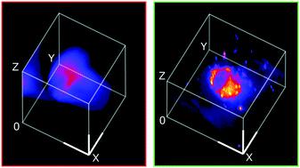
Moldable, transparent polydimethylsiloxane (PDMS) elastomer microdevices enable a broad range of complex studies of three-dimensional cellular networks in their microenvironment in vitro. However, the uneven distribution of refractive index change, external to PDMS devices and internally in the sample chamber, creates a significant optical path difference (OPD) that distorts the light sheet beam and so restricts diffraction limited performance. We experimentally showed that an OPD of 120 μm results in the broadening of the lateral point spread function by over 4-fold. In this paper, we demonstrate steps to adapt a commercial inverted selective plane illumination microscope (iSPIM) and remove the OPD so as to achieve sub-micrometer imaging ranging from 0.6 ± 0.04 μm to 0.91 ± 0.03 μm of a fluorescence biological sample suspended in regular saline (RI ≈1.34) enclosed in 1.2 to 2 mm thick micromolded PDMS microdevices. We have proven that the removal of the OPD from the external PDMS layer by refractive index (RI) matching with a readily accessible, inexpensive sucrose solution is critical to achieve a >3-fold imaging resolution improvement. To monitor the RI matching process, a single-mode fiber (SMF) illuminator was integrated into the iSPIM. To remove the OPD inside the PDMS channel, we used an electrically tunable lens (ETL) that par-focuses the light sheet beam with the detection objective lens and so minimised axial distortions to attain sub-micrometer imaging resolution. We termed this new light sheet imaging protocol as modified inverted selective plane illumination microscopy (m-iSPIM). Using the high spatial–temporal 3D imaging of m-iSPIM, we experimentally captured single platelet (≈2 μm) recruitment to a platelet aggregate (22.5 μm × 22.5 μm × 6 μm) under flow at a 150 μm depth within a microfluidic channel. m-iSPIM paves the way for the application of light sheet imaging to a wide range of 3D biological models in microfluidic devices which recapitulate features of the physiological microenvironment and elucidate subcellular responses.
中文翻译:

在聚二甲基硅氧烷软光刻设备中用于亚微米成像分辨率的改进的倒置选择性平面照明显微镜。
可模制的透明聚二甲基硅氧烷(PDMS)弹性体微器件可在其微环境中进行广泛的三维细胞网络复杂研究。但是,PDMS装置外部和样品室内部的折射率变化的不均匀分布会产生显着的光程差(OPD),从而使光束变形,从而限制了衍射受限的性能。我们通过实验表明,120μm的OPD会导致横向点扩展功能变宽4倍以上。在本文中,我们演示了采用商用倒置选择性平面照明显微镜(iSPIM)并去除OPD的步骤,以实现从0.6±0.04μm到0.91±0.03μm的悬浮在常规环境中的荧光生物样品的亚微米成像盐溶液(RI≈1.34)封装在1.2至2毫米厚的微成型PDMS微型设备中。我们已经证明,通过折射率(RI)与易于获得的廉价蔗糖溶液匹配来从外部PDMS层中去除OPD对于实现> 3倍的成像分辨率提高至关重要。为了监视RI匹配过程,将单模光纤(SMF)照明器集成到iSPIM中。为了去除PDMS通道内的OPD,我们使用了一个电可调透镜(ETL),该透镜将光束与检测物镜齐焦,从而最大程度地减小了轴向畸变,从而获得了亚微米级的成像分辨率。我们称这种新的光片成像协议为改良的倒置选择性平面照明显微镜(m-iSPIM)。使用m-iSPIM的高时空3D成像,我们实验性地捕获了单个血小板(≈2μm)募集到血小板聚集体(22.5μm×22)。5μm×6μm)在微流体通道中以150μm的深度流动。m-iSPIM为将光片成像应用到微流控设备中的各种3D生物学模型铺平了道路,这些设备概括了生理微环境的特征并阐明了亚细胞反应。
更新日期:2020-09-18
中文翻译:
在聚二甲基硅氧烷软光刻设备中用于亚微米成像分辨率的改进的倒置选择性平面照明显微镜。
可模制的透明聚二甲基硅氧烷(PDMS)弹性体微器件可在其微环境中进行广泛的三维细胞网络复杂研究。但是,PDMS装置外部和样品室内部的折射率变化的不均匀分布会产生显着的光程差(OPD),从而使光束变形,从而限制了衍射受限的性能。我们通过实验表明,120μm的OPD会导致横向点扩展功能变宽4倍以上。在本文中,我们演示了采用商用倒置选择性平面照明显微镜(iSPIM)并去除OPD的步骤,以实现从0.6±0.04μm到0.91±0.03μm的悬浮在常规环境中的荧光生物样品的亚微米成像盐溶液(RI≈1.34)封装在1.2至2毫米厚的微成型PDMS微型设备中。我们已经证明,通过折射率(RI)与易于获得的廉价蔗糖溶液匹配来从外部PDMS层中去除OPD对于实现> 3倍的成像分辨率提高至关重要。为了监视RI匹配过程,将单模光纤(SMF)照明器集成到iSPIM中。为了去除PDMS通道内的OPD,我们使用了一个电可调透镜(ETL),该透镜将光束与检测物镜齐焦,从而最大程度地减小了轴向畸变,从而获得了亚微米级的成像分辨率。我们称这种新的光片成像协议为改良的倒置选择性平面照明显微镜(m-iSPIM)。使用m-iSPIM的高时空3D成像,我们实验性地捕获了单个血小板(≈2μm)募集到血小板聚集体(22.5μm×22)。5μm×6μm)在微流体通道中以150μm的深度流动。m-iSPIM为将光片成像应用到微流控设备中的各种3D生物学模型铺平了道路,这些设备概括了生理微环境的特征并阐明了亚细胞反应。


 京公网安备 11010802027423号