Journal of Neuroscience Methods ( IF 2.7 ) Pub Date : 2020-09-05 , DOI: 10.1016/j.jneumeth.2020.108926 Zhenxiong Wang 1 , Mehran Shaghaghi 2 , Shun Zhang 3 , Guiling Zhang 3 , Yiran Zhou 3 , Di Wu 3 , Zhuoli Zhang 4 , Wenzhen Zhu 3 , Kejia Cai 2
|
Background
To map and quantify the proton exchange rate () of brain tissues using improved omega plots in ischemic stroke patients and to investigate whether can serve as a potential endogenous surrogate imaging biomarker for detecting the metabolic state and the pathologic changes due to ischemic stroke.
New Method
Three sets of Z-spectra were acquired from seventeen ischemic stroke patients using a spin echo-echo planar imaging sequence with pre-saturation chemical exchange saturation transfer (CEST) pulse at of 1.5, 2.5, and 3.5 , respectively. Pixel-wise was calculated from improved omega plot of water direct saturation (DS)-removed Z-spectral signals.
Results
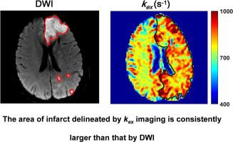
The derived maps can differentiate infarcts from contralateral normal brain tissues with significantly increased signal (893 ± 52 vs. 739 ± 34 , P < 0.001).
Comparison with Existing Method(s)
The maps were found to be different from conventional contrasts from diffusion-weighted imaging (DWI), CEST, and semi-solid magnetization transfer (MT) MRI. In brief, MRI showed larger lesion areas than DWI with different degrees and different lesion contrast compared to CEST and MT.
Conclusions
In this preliminary translational research, the MRI based on DS-removed omega plots has been demonstrated for in vivo imaging of clinical ischemic stroke patients. As a noninvasive and unique MRI contrast, MRI at 3 T may serve as a potential surrogate imaging biomarker for the metabolic changes of stroke and help for monitoring the evolution and the treatment of stroke.
中文翻译:
新型质子交换率 MRI 在缺血性中风患者的大脑中呈现独特的对比。
背景
映射和量化质子交换率() 使用改进的 omega 图在缺血性中风患者中分析脑组织,并调查是否 可作为潜在的内源性替代成像生物标志物,用于检测缺血性卒中引起的代谢状态和病理变化。
新方法
使用具有预饱和化学交换饱和转移 (CEST) 脉冲的自旋回波回波平面成像序列从 17 名缺血性中风患者获得三组 Z 谱。 1.5、2.5 和 3.5 , 分别。像素级 由水直接饱和度 (DS) 去除的 Z 光谱信号的改进 omega 图计算。
结果
派生的 图可以区分梗死和对侧正常脑组织,信号显着增加(893±52 与. 739±34, P < 0.001)。
与现有方法的比较
这 发现地图不同于来自扩散加权成像 (DWI)、CEST 和半固体磁化转移 (MT) MRI 的传统对比。简单来说, 与 CEST 和 MT 相比,MRI 显示比 DWI 更大的病变区域,具有不同程度和不同的病变对比度。
结论
在这项初步的转化研究中, 基于去除 DS 的 omega 图的 MRI 已被证明可用于临床缺血性中风患者的体内成像。作为一种无创且独特的 MRI 对比, 3 T MRI 可作为脑卒中代谢变化的潜在替代成像生物标志物,有助于监测脑卒中的演变和治疗。


 京公网安备 11010802027423号