Annals of Anatomy ( IF 2.0 ) Pub Date : 2020-09-06 , DOI: 10.1016/j.aanat.2020.151593 Azzat Al-Redouan 1 , Keiv Holding 1 , David Kachlik 1
|
Background
Suprascapular nerve (SN) entrapment syndrome accounts for 1-2% of all shoulder pain. The SN travels within a space between the suprascapular notch (SSN) and the spinoglenoid notch (SGN).
Purpose
To report a detailed topographical study of the suprascapular canal (SSC) and ultimately sort the different types of SN entrapment by its anatomical localization within the canal.
Basic procedures
Observational study on 30 free dissected limbs of formaldehyde-fixed cadavers. The SN and vessels were traced as they passed through the SSC and the boundaries of the SSC were observed and documented. The SSC was then exposed by reflecting away the bordering muscles. Dimensions of the SSC as well as parameters of the SSN and SGN were measured using a digital caliper. Finally, a thorough literature review was made to survey the SN entrapment occurrence by site.
Main findings
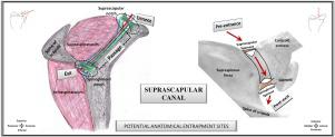
The SSC is situated in the spinoglenoid fossa, has an average width of 13 mm, and runs underneath the supraspinatus muscle with an average distance of 25 mm between the SSN and SGN sloping in an infero-postero-lateral direction. The first segment represents the SSC entrance site and is composed of two spaces: osteofibrous and musculofibrous. The second segment is bordered by the supraspinatus muscle fascia, lateral margin of the supraspinous fossa, glenohumeral joint capsule, and the bony surface of the scapula (spinoglenoid fossa). This represents the SSC passage site. The third segment represents the SSC exit site around the spinoacromial arch at the SGN.
Principal conclusions
The SSC is defined as an osteofibrous canal running between the SSN and SGN enclosed by the supraspinatus fascia. It is anatomically composed of three segments: an entrance, a passage, and an exit. The distal SN passes through the SSC via five intervals that correspond to five potential sites of anatomical nerve entrapment: at the pre-entrance site, entrance site, passage site, exit site, and post-exit site. Each of those sites was found to be associated with specific causes and forms of entrapment.
中文翻译:
“肩胛上管”:解剖和地形描述及其在卡压综合征中的临床意义。
背景
肩胛上神经 (SN) 卡压综合征占所有肩痛的 1-2%。SN 在肩胛上切迹 (SSN) 和棘突切迹 (SGN) 之间的空间内移动。
目的
报告对肩胛上管 (SSC) 的详细地形研究,并最终根据其在管内的解剖定位对不同类型的 SN 诱捕进行分类。
基本程序
30具甲醛固定尸体游离解剖肢体的观察研究。SN 和船只在通过 SSC 时被追踪,SSC 的边界被观察和记录。然后通过反射远离边界的肌肉来暴露 SSC。使用数显卡尺测量 SSC 的尺寸以及 SSN 和 SGN 的参数。最后,进行了彻底的文献回顾,以按站点调查 SN 诱捕事件。
主要发现
SSC 位于棘突窝,平均宽度为 13 毫米,在冈上肌下方运行,SSN 和 SGN 之间的平均距离为 25 毫米,呈下-后-外侧方向倾斜。第一段代表 SSC 入口部位,由两个空间组成:骨纤维和肌肉纤维。第二节以冈上肌筋膜、冈上窝外侧缘、盂肱关节囊和肩胛骨(棘突窝)为界。这代表 SSC 通道站点。第三段代表 SGN 脊柱肩峰弓周围的 SSC 出口部位。
主要结论
SSC 被定义为在 SSN 和 SGN 之间运行的骨纤维管,被冈上肌筋膜包围。它在解剖学上由三部分组成:入口、通道和出口。远端 SN 通过五个间隔穿过 SSC,这些间隔对应于解剖神经卡压的五个潜在部位:在入口前部位、入口部位、通道部位、出口部位和出口后部位。发现这些地点中的每一个都与诱捕的具体原因和形式有关。


 京公网安备 11010802027423号