Nano Research ( IF 9.9 ) Pub Date : 2020-08-10 , DOI: 10.1007/s12274-020-2982-7 Mo Chen , Sijia Feng , Yimeng Yang , Yunxia Li , Jian Zhang , Shiyi Chen , Jun Chen
|
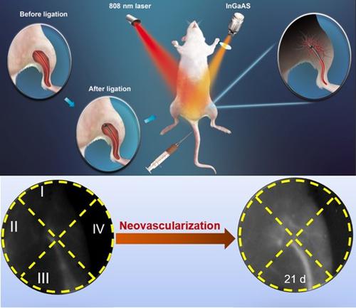
The in vivo spatio-temporal patterns of neovascularization are still poorly understood because it is limited to multi-scale techniques from the cellular level to living animal level. Owing to deep tissue-penetration and zero autofluorescence background, the second near-infrared (NIR-II, 1,000–1,700 nm) fluorescence imaging recently shows promise in breaking through this dilemma by dynamically tracking the pathophysiological process of neovascularization in vivo. Here, NIR-II fluorescence imaging was recruited for monitoring blood vessels in order to visualize the vascular injury and quantitively assess neovascularization in mouse models of acute skeleton muscle contusion and hindlimb ischemia. The temporal analysis of real-time NIR-II fluorescence intensity demonstrated that the blood flow perfusion of ischemia area was able to rapidly restore to 96% of pre-ischemic state within one week. Moreover, the spatial analysis revealed that the lower and outer quadrants of ischemia area in the mouse model of hindlimb ischemia always had relatively high blood flow perfusion compared with other quadrants during three weeks post-ischemia, and even exceeded pre-ischemic quantity at 21 days post-ischemia. In conclusion, this in vivo imaging technique has significant potential utility for studying the spatio-temporal patterns of neovascularization in vivo.
中文翻译:
通过NIR-II荧光成像跟踪体内新血管形成的时空模式
的体内新血管形成的时空图案仍知之甚少,因为它是从细胞水平对活的动物水平限于多尺度技术。由于深层组织穿透和零自发荧光背景,第二次近红外(NIR-II,1,000-1,700 nm)荧光成像最近显示了有望通过动态跟踪体内新血管形成的病理生理过程突破这一难题的希望。。在这里,NIR-II荧光成像被招募来监测血管,以可视化血管损伤并定量评估急性骨骼肌挫伤和后肢缺血的小鼠模型中的新血管形成。实时NIR-II荧光强度的时间分析表明,缺血区域的血流灌注能够在一周内迅速恢复至缺血前状态的96%。此外,空间分析显示,在缺血后三周内,后肢缺血小鼠模型中缺血区的下象限和外象限始终具有相对较高的血流灌注,甚至在第21天超过了缺血前量缺血后。总之,这种体内成像技术对于研究体内新血管形成的时空模式具有重要的潜在实用性。


 京公网安备 11010802027423号