当前位置:
X-MOL 学术
›
J. Mater. Chem. B
›
论文详情
Our official English website, www.x-mol.net, welcomes your
feedback! (Note: you will need to create a separate account there.)
Superhydrophobic bowl-like SERS substrates patterned from CMOS sensors for extracellular vesicle characterization
Journal of Materials Chemistry B ( IF 6.1 ) Pub Date : 2020-09-02 , DOI: 10.1039/d0tb00889c Sorina Suarasan 1, 2, 3, 4 , Juanjuan Liu 1, 2, 3, 4 , Meruyert Imanbekova 1, 2, 3, 4 , Tatu Rojalin 5, 6, 7, 8 , Silvia Hilt 6, 7, 8, 9 , John C. Voss 6, 7, 8, 9 , Sebastian Wachsmann-Hogiu 1, 2, 3, 4
Journal of Materials Chemistry B ( IF 6.1 ) Pub Date : 2020-09-02 , DOI: 10.1039/d0tb00889c Sorina Suarasan 1, 2, 3, 4 , Juanjuan Liu 1, 2, 3, 4 , Meruyert Imanbekova 1, 2, 3, 4 , Tatu Rojalin 5, 6, 7, 8 , Silvia Hilt 6, 7, 8, 9 , John C. Voss 6, 7, 8, 9 , Sebastian Wachsmann-Hogiu 1, 2, 3, 4
Affiliation
|
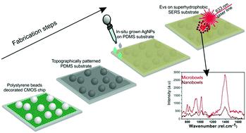
Using a regular CMOS sensor as a template, we are able to fabricate a simple but highly effective superhydrophobic SERS substrate. Specifically, we decorated the microlens layer of the sensor with 7 μm polystyrene beads to obtain a PDMS patterned replica. The process resulted in a uniform pattern of voids in the PDMS (denoted nanobowls) that are intercalated with a few larger voids (denoted here microbowls). The voids act as superhydrophobic substrates with analyte concentration capabilities in bigger bowl-like structures. Silver nanoparticles were directly grown on the patterned PDMS substrate inside both the nano- and microbowls, and serve as strong electromagnetic field enhancers for the SERS substrate. After systematic characterization of the fabricated SERS substrate by atomic force microscopy and scanning electron microscopy, we demonstrated its SERS performance using 4-aminothiophenol as a reporter molecule. Finally, we employed this innovative substrate to concentrate and analyze extracellular vesicles (EVs) isolated from an MC65 neural cell line in an ultralow sample volume. This substrate can be further exploited for the investigation of various EV biomarkers for early diagnosis of different diseases using liquid biopsy.
中文翻译:

利用CMOS传感器图案化的超疏水碗状SERS基质,用于细胞外囊泡表征
使用常规的CMOS传感器作为模板,我们能够制造出简单但高效的超疏水SERS基板。具体而言,我们用7μm聚苯乙烯珠装饰了传感器的微透镜层,以获得PDMS图案化的副本。该过程导致PDMS(表示为纳米碗)中的空隙均匀分布,并插入了一些较大的空隙(此处称为微碗)。空隙充当较大的碗状结构中具有分析物浓缩能力的超疏水性底物。银纳米颗粒直接生长在纳米碗和微碗内部的带图案的PDMS衬底上,并充当SERS衬底的强电磁场增强剂。在通过原子力显微镜和扫描电子显微镜对制成的SERS基板进行系统表征后,我们使用4-氨基硫酚作为报告分子证明了其SERS性能。最后,我们采用了这种创新的底物,以超低的样品量浓缩和分析了从MC65神经细胞系分离出的细胞外囊泡(EV)。该底物可进一步用于研究各种EV生物标志物,以便使用液体活检来早期诊断不同疾病。
更新日期:2020-10-07
中文翻译:
利用CMOS传感器图案化的超疏水碗状SERS基质,用于细胞外囊泡表征
使用常规的CMOS传感器作为模板,我们能够制造出简单但高效的超疏水SERS基板。具体而言,我们用7μm聚苯乙烯珠装饰了传感器的微透镜层,以获得PDMS图案化的副本。该过程导致PDMS(表示为纳米碗)中的空隙均匀分布,并插入了一些较大的空隙(此处称为微碗)。空隙充当较大的碗状结构中具有分析物浓缩能力的超疏水性底物。银纳米颗粒直接生长在纳米碗和微碗内部的带图案的PDMS衬底上,并充当SERS衬底的强电磁场增强剂。在通过原子力显微镜和扫描电子显微镜对制成的SERS基板进行系统表征后,我们使用4-氨基硫酚作为报告分子证明了其SERS性能。最后,我们采用了这种创新的底物,以超低的样品量浓缩和分析了从MC65神经细胞系分离出的细胞外囊泡(EV)。该底物可进一步用于研究各种EV生物标志物,以便使用液体活检来早期诊断不同疾病。


 京公网安备 11010802027423号