Annals of Anatomy ( IF 2.0 ) Pub Date : 2020-09-02 , DOI: 10.1016/j.aanat.2020.151588 Adam Ostrzenski 1
|
Background
To describe the BV anatomy in detail, to compare previous BV descriptions and illustrations to the current study’s findings and photograms, to show the BV topographic relation of the BV to the urethral meatus, to document the BV anatomy using photograms.
Methods
Ten fresh human female adult cadavers were used. Stratum-by-stratum anatomical dissections in sagittal, transverse, and coronal planes were performed. The BV was dissected-off from the original location of the posterior-distal vaginal wall and the anterior anal wall.
Results
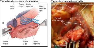
The BV was located within the posterior-distal vagina and composed of two vertical legs, which fused to one another. The inferior pars intermedia fused both descending legs to the anterior-proximal perineal urethral wall, and BV embraced the anterior-proximal urethra. The superior pars intermedia connects the BV to the posterior-distal clitoral body. The BV legs traversed parallel to and aside from the vaginal introitus and the lateral urethra and not crossing the anterior-distal urethra. The tile-end was a tapered end which terminates in the vicinity of Bartholin glands. Laterally, the BV legs outspread to the medial labia minora and attach to the ischiopubic ramus. The anatomical site-specific defect (s) occurs within the BV.
Conclusions
The present study resolves the BV anatomical controversy and shows that the BV runs parallel to and aside from the anterior-distal urethra and the BV. The site-specific defect(s) can occur within the BV. This study provides important information for anatomy educators and surgeons.
中文翻译:
球茎前庭的解剖:尸体研究。
背景
详细描述 BV 解剖结构,将以前的 BV 描述和插图与当前研究的结果和照片进行比较,显示 BV 与尿道口的 BV 地形关系,使用照片记录 BV 解剖结构。
方法
使用了十具新鲜的人类女性成年尸体。在矢状面、横向和冠状面进行逐层解剖解剖。从阴道后壁和肛门前壁的原始位置切下 BV。
结果
BV 位于阴道后部远端,由两条相互融合的垂直腿组成。下中间部分将两条降支腿与会阴前近端尿道壁融合,BV 包围前近端尿道。上中部将 BV 连接到后-远端阴蒂体。BV 腿平行于阴道口和外侧尿道并在其旁边横穿,而不穿过前-远侧尿道。瓦端是在前庭大腺附近终止的锥形端。从侧面看,BV 腿延伸到内侧小阴唇并附着在坐骨耻骨支上。解剖部位特异性缺陷发生在 BV 内。
结论
本研究解决了 BV 解剖学上的争议,并表明 BV 与前远端尿道和 BV 平行并远离。特定于站点的缺陷可能发生在 BV 内。这项研究为解剖学教育者和外科医生提供了重要信息。


 京公网安备 11010802027423号