Bioorganic Chemistry ( IF 4.5 ) Pub Date : 2020-09-02 , DOI: 10.1016/j.bioorg.2020.104267 Xin-Yue Shang 1 , Rui Guo 1 , Xiao-Qi Yu 1 , Bin Lin 2 , Xiao-Xiao Huang 1 , Guo-Dong Yao 1 , Shao-Jiang Song 1
|
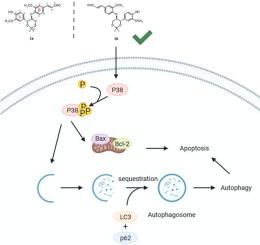
Crataegus pinnatifida has been famous for its nutritional purpose. However, systematic investigation on the bioactive constituents is still lacking, although this fruit has been reported for its cytotoxic effect before. In this study, two pairs of new lignan enantiomers (1a/1b, 2a/2b), which isolated using chiral chromatographic column from the fruits of C. pinnatifida were studied. The absolute configurations of enantiomers were determined by comparison between the experimental electronic circular dichroism (ECD) and calculated ECD spectra. Among them, 1a/1b exhibited a better cytotoxic effect in hepatocellular carcinoma Hep3B cells with an IC50 value of 34.97 ± 2.74 and 17.42 ± 0.71 μM, respectively. In addition, 1b induced much more apoptotic, autophagic cells than 1a in Hep3B cells. Furthermore, the underlying mechanism was demonstrated that p38 activation could promote 1b-induced apoptosis and autophagy. Moreover, 1b-induced apoptosis was significantly decreased in the presence of autophagic inhibitor Bafilomycin A1 (Baf A1), suggesting that the induction of autophagy enhanced apoptotic cell death in 1b-treated cells. In general, these findings provide a valuable basis for further understanding the effect of 8-O-4′ lignans in C. pinnatifida on cytotoxic effect.
中文翻译:
对虾Cretaegus pinnatifida的对映体8-O-4'型新木脂素通过Hep3B细胞的凋亡和自噬作用表现出细胞毒性作用。
Crataegus pinnatifida以其营养目的而闻名。但是,尽管以前已经报道过这种水果具有细胞毒性作用,但仍缺乏对生物活性成分的系统研究。在这项研究中,研究了两对使用手性色谱柱从C. pinnatifida果实中分离的新的木脂素对映体(1a / 1b,2a / 2b)。对映体的绝对构型是通过比较实验电子圆二色性(ECD)和计算出的ECD光谱确定的。其中1a / 1b在具有IC的肝细胞癌Hep3B细胞中表现出更好的细胞毒性作用50值分别为34.97±2.74和17.42±0.71μM。另外,在Hep3B细胞中,1b诱导的凋亡自噬细胞比1a多得多。此外,潜在的机制被证明p38激活可以促进1b诱导的细胞凋亡和自噬。此外,在自噬抑制剂巴氟霉素A1(Baf A1)存在下,1b诱导的细胞凋亡显着减少,这表明自噬的诱导增强了1b处理细胞的凋亡。通常,这些发现为进一步了解pinnatifida中的8- O -4'木脂素对细胞毒性作用的影响提供了有价值的基础。


 京公网安备 11010802027423号