Our official English website, www.x-mol.net, welcomes your feedback! (Note: you will need to create a separate account there.)
Microfluidic-prepared, monodisperse, X-ray-visible, embolic microspheres for non-oncological embolization applications.
Lab on a Chip ( IF 6.1 ) Pub Date : 2020-09-01 , DOI: 10.1039/d0lc00098a Cyrus W Beh 1 , Yingli Fu , Clifford R Weiss , Charles Hu , Aravind Arepally , Hai-Quan Mao , Tza-Huei Wang , Dara L Kraitchman
Lab on a Chip ( IF 6.1 ) Pub Date : 2020-09-01 , DOI: 10.1039/d0lc00098a Cyrus W Beh 1 , Yingli Fu , Clifford R Weiss , Charles Hu , Aravind Arepally , Hai-Quan Mao , Tza-Huei Wang , Dara L Kraitchman
Affiliation
|
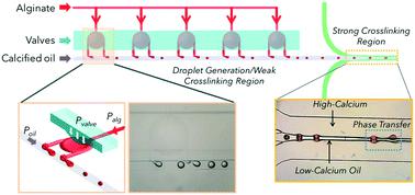
Embolotherapy using particle embolics is normally performed with exogenous contrast to assist in visualization. However, the exact location of the embolics cannot be identified after contrast washout. We developed a novel, pseudo-check valve-integrated microfluidic device, that partitions barium- impregnated alginate from crosslinking solution, thereby preventing nozzle failure. This enables rapid and continuous generation of inherently X-ray-visible embolic microspheres (XEMs) with uniform size. The XEMs are visible under clinical X-ray and cone beam CT both in vitro and in vivo. In particular, we demonstrated the embolization properties of these XEMs in large animals, performing direct intra- and post-procedural assessment of embolic delivery. The persistent radiopacity of these XEMs enables real-time evaluation of embolization precision and offers great promise for non-invasive follow-up examination without exogenous contrast. We also demonstrated that bariatric arterial embolization with XEMs significantly suppresses weight gain in swine, as an example of a non-oncological application of embolotherapy.
中文翻译:

用于非肿瘤栓塞应用的微流体制备、单分散、X 射线可见的栓塞微球。
使用颗粒栓塞的栓塞疗法通常使用外源造影剂进行,以帮助可视化。然而,在造影剂清除后无法确定栓塞的确切位置。我们开发了一种新型的伪止回阀集成微流体装置,可将浸渍海藻酸钡的溶液与交联溶液隔开,从而防止喷嘴故障。这使得能够快速、连续地生成具有均匀大小的固有 X 射线可见栓塞微球 (XEM)。XEM在体外和体内的临床 X 射线和锥形束 CT 下都可见. 特别是,我们展示了这些 XEM 在大型动物中的栓塞特性,对栓塞递送进行了直接的程序内和程序后评估。这些 XEM 的持久辐射不透性使得能够实时评估栓塞精度,并为无需外源造影剂的非侵入性后续检查提供了巨大希望。我们还证明,使用 XEM 进行减肥动脉栓塞可显着抑制猪的体重增加,作为栓塞疗法非肿瘤应用的一个例子。
更新日期:2020-09-29
中文翻译:
用于非肿瘤栓塞应用的微流体制备、单分散、X 射线可见的栓塞微球。
使用颗粒栓塞的栓塞疗法通常使用外源造影剂进行,以帮助可视化。然而,在造影剂清除后无法确定栓塞的确切位置。我们开发了一种新型的伪止回阀集成微流体装置,可将浸渍海藻酸钡的溶液与交联溶液隔开,从而防止喷嘴故障。这使得能够快速、连续地生成具有均匀大小的固有 X 射线可见栓塞微球 (XEM)。XEM在体外和体内的临床 X 射线和锥形束 CT 下都可见. 特别是,我们展示了这些 XEM 在大型动物中的栓塞特性,对栓塞递送进行了直接的程序内和程序后评估。这些 XEM 的持久辐射不透性使得能够实时评估栓塞精度,并为无需外源造影剂的非侵入性后续检查提供了巨大希望。我们还证明,使用 XEM 进行减肥动脉栓塞可显着抑制猪的体重增加,作为栓塞疗法非肿瘤应用的一个例子。


 京公网安备 11010802027423号