当前位置:
X-MOL 学术
›
Brain Pathol.
›
论文详情
Our official English website, www.x-mol.net, welcomes your feedback! (Note: you will need to create a separate account there.)
Distinct changes in all major components of the neurovascular unit across different neuropathological stages of Alzheimer's disease.
Brain Pathology ( IF 6.4 ) Pub Date : 2020-08-31 , DOI: 10.1111/bpa.12895 Tunahan Kirabali 1 , Ruslan Rust 1 , Serena Rigotti 1, 2 , Alessandro Siccoli 1, 3 , Roger M Nitsch 1, 4 , Luka Kulic 1, 5
Brain Pathology ( IF 6.4 ) Pub Date : 2020-08-31 , DOI: 10.1111/bpa.12895 Tunahan Kirabali 1 , Ruslan Rust 1 , Serena Rigotti 1, 2 , Alessandro Siccoli 1, 3 , Roger M Nitsch 1, 4 , Luka Kulic 1, 5
Affiliation
|
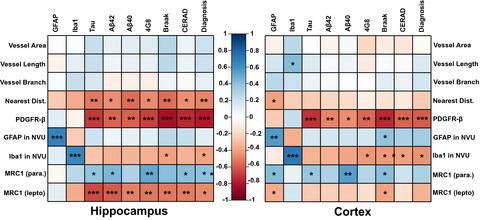
In the brain capillaries, endothelial cells, pericytes, astrocytes and microglia form a structural and functional complex called neurovascular unit (NVU) which is critically involved in maintaining neuronal homeostasis. In the present study, we applied a comprehensive immunohistochemical approach to investigate the structural alterations in the NVU across different Alzheimer's disease (AD) neuropathological stages. Post‐mortem human cortical and hippocampal samples derived from AD patients and non‐demented elderly control individuals were immunostained using a panel of markers representing specific components of the NVU including Collagen IV (basement membrane), PDGFR‐β (pericytes), GFAP (astrocytes), Iba1 (microglia), MRC1 (perivascular macrophages) and lectin as an endothelial cell label. Astrocytes (GFAP) and microglia (Iba1) were quantified both in the whole visual‐field and specifically within the NVU, and the sample set was additionally analyzed using anti‐tau (AT8) and three different anti‐Aβ (clones G2‐10, G2‐11, 4G8) antibodies. Analyses of lectin labeled sections showed an altered vascular distribution in AD patients as revealed by a reduced nearest distance between capillaries. Within the NVU, a Braak‐stage dependent reduction in pericyte coverage was identified as the earliest structural alteration during AD progression. In comparison to non‐demented elderly controls, AD patients showed a significantly higher astrocyte coverage within the NVU, which was paralleled by a reduced microglial coverage around capillaries. Assessment of perivascular macrophages moreover demonstrated a relocation of these cells from leptomeningeal arteries to penetrating parenchymal vessels in AD patients. Collectively, the results of our study represent a comprehensive first in‐depth analysis of AD‐related structural changes in the NVU and suggest distinct alterations in all components of the NVU during AD progression.
中文翻译:

阿尔茨海默病不同神经病理学阶段神经血管单元所有主要成分的明显变化。
在脑毛细血管中,内皮细胞、周细胞、星形胶质细胞和小胶质细胞形成称为神经血管单元 (NVU) 的结构和功能复合体,其在维持神经元稳态方面发挥着重要作用。在本研究中,我们应用全面的免疫组织化学方法来研究不同阿尔茨海默病 (AD) 神经病理学阶段的 NVU 结构变化。来自 AD 患者和非痴呆老年对照个体的死后人类皮质和海马样本使用一组代表 NVU 特定成分的标记物进行免疫染色,包括胶原蛋白 IV(基底膜)、PDGFR-β(周细胞)、GFAP(星形胶质细胞) )、Iba1(小胶质细胞)、MRC1(血管周围巨噬细胞)和凝集素作为内皮细胞标记。星形胶质细胞 (GFAP) 和小胶质细胞 (Iba1) 在整个视野中,特别是在 NVU 内进行量化,并且使用抗 tau (AT8) 和三种不同的抗 Aβ(克隆 G2-10, G2-11, 4G8) 抗体。凝集素标记切片的分析显示 AD 患者的血管分布发生了改变,这通过毛细血管之间的最近距离减小来揭示。在 NVU 中,Braak 阶段依赖性周细胞覆盖减少被确定为 AD 进展过程中最早的结构改变。与非痴呆的老年对照相比,AD 患者在 NVU 内显示出明显更高的星形胶质细胞覆盖率,同时毛细血管周围的小胶质细胞覆盖率降低。此外,血管周围巨噬细胞的评估表明,这些细胞从软脑膜动脉迁移到 AD 患者的穿透实质血管。总的来说,我们的研究结果代表了对 NVU 中 AD 相关结构变化的首次全面深入分析,并表明 AD 进展过程中 NVU 的所有组成部分都有明显的改变。
更新日期:2020-08-31
中文翻译:
阿尔茨海默病不同神经病理学阶段神经血管单元所有主要成分的明显变化。
在脑毛细血管中,内皮细胞、周细胞、星形胶质细胞和小胶质细胞形成称为神经血管单元 (NVU) 的结构和功能复合体,其在维持神经元稳态方面发挥着重要作用。在本研究中,我们应用全面的免疫组织化学方法来研究不同阿尔茨海默病 (AD) 神经病理学阶段的 NVU 结构变化。来自 AD 患者和非痴呆老年对照个体的死后人类皮质和海马样本使用一组代表 NVU 特定成分的标记物进行免疫染色,包括胶原蛋白 IV(基底膜)、PDGFR-β(周细胞)、GFAP(星形胶质细胞) )、Iba1(小胶质细胞)、MRC1(血管周围巨噬细胞)和凝集素作为内皮细胞标记。星形胶质细胞 (GFAP) 和小胶质细胞 (Iba1) 在整个视野中,特别是在 NVU 内进行量化,并且使用抗 tau (AT8) 和三种不同的抗 Aβ(克隆 G2-10, G2-11, 4G8) 抗体。凝集素标记切片的分析显示 AD 患者的血管分布发生了改变,这通过毛细血管之间的最近距离减小来揭示。在 NVU 中,Braak 阶段依赖性周细胞覆盖减少被确定为 AD 进展过程中最早的结构改变。与非痴呆的老年对照相比,AD 患者在 NVU 内显示出明显更高的星形胶质细胞覆盖率,同时毛细血管周围的小胶质细胞覆盖率降低。此外,血管周围巨噬细胞的评估表明,这些细胞从软脑膜动脉迁移到 AD 患者的穿透实质血管。总的来说,我们的研究结果代表了对 NVU 中 AD 相关结构变化的首次全面深入分析,并表明 AD 进展过程中 NVU 的所有组成部分都有明显的改变。


 京公网安备 11010802027423号