Our official English website, www.x-mol.net, welcomes your
feedback! (Note: you will need to create a separate account there.)
Plasmonic Nanoparticle-Based Digital Cytometry to Quantify MUC16 Binding on the Surface of Leukocytes in Ovarian Cancer.
ACS Sensors ( IF 8.2 ) Pub Date : 2020-08-27 , DOI: 10.1021/acssensors.0c00567 Sinyoung Jeong 1 , Germán González 2 , Alexander Ho 1 , Nicholas Nowell 2 , Lauren A Austin 1 , Jawad Hoballah 2 , Fatima Mubarak 2 , Arvinder Kapur 3 , Manish S Patankar 3 , Daniel W Cramer 4, 5 , Petra Krauledat 2 , W Peter Hansen 2 , Conor L Evans 1, 6
ACS Sensors ( IF 8.2 ) Pub Date : 2020-08-27 , DOI: 10.1021/acssensors.0c00567 Sinyoung Jeong 1 , Germán González 2 , Alexander Ho 1 , Nicholas Nowell 2 , Lauren A Austin 1 , Jawad Hoballah 2 , Fatima Mubarak 2 , Arvinder Kapur 3 , Manish S Patankar 3 , Daniel W Cramer 4, 5 , Petra Krauledat 2 , W Peter Hansen 2 , Conor L Evans 1, 6
Affiliation
|
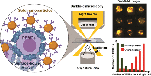
Although levels of the circulating ovarian cancer marker (CA125) can distinguish ovarian masses that are likely to be malignant and correlate with severity of disease, serum CA125 has not proved useful in general population screening. Recently, cell culture studies have indicated that MUC16 may bind to the Siglec-9 receptor on natural killer (NK) cells where it downregulates the cytotoxicity of NK cells, allowing ovarian cancer cells to evade immune surveillance. We present evidence that the presence of MUC16 can be locally visualized and imaged on the surface of peripheral blood mononuclear cells (PBMCs) in ovarian cancer via a novel “digital” cytometry technique that incorporates: (i) OC125 monoclonal antibody-conjugated gold nanoparticles as optical nanoprobes, (ii) a high contrast dark-field microscopy system to detect PBMC-bound gold nanoparticles, and (iii) a computational algorithm for automatic counting of these nanoparticles to estimate the quantity of surface-bound MUC16. The quantitative detection of our technique was successfully demonstrated by discriminating clones of the ovarian cancer cell line, OVCAR3, based on low, intermediate, and high expression levels of MUC16. Additionally, PBMC surface-bound MUC16 was tracked in an ovarian cancer patient over a 17 month period; the results suggest that the binding of MUC16 on the surface of immune cells may play an early indicator for recurrent metastasis 6 months before computational tomography-based clinical diagnosis. We also demonstrate that the levels of surface-bound MUC16 on PBMCs from five ovarian cancer patients were greater than those from five healthy controls.
中文翻译:

基于等离子体纳米颗粒的数字细胞计数法可量化卵巢癌白细胞表面 MUC16 的结合。
尽管循环卵巢癌标志物 (CA125) 的水平可以区分可能是恶性的卵巢肿块并与疾病严重程度相关,但血清 CA125 尚未证明在一般人群筛查中有用。最近,细胞培养研究表明,MUC16 可能与自然杀伤 (NK) 细胞上的 Siglec-9 受体结合,下调 NK 细胞的细胞毒性,从而使卵巢癌细胞逃避免疫监视。我们提供的证据表明,MUC16 的存在可以通过一种新颖的“数字”细胞计数技术在卵巢癌的外周血单核细胞 (PBMC) 表面进行局部可视化和成像,该技术包括:(i) OC125 单克隆抗体缀合的金纳米粒子作为光学纳米探针,(ii) 用于检测 PBMC 结合金纳米粒子的高对比度暗视野显微镜系统,以及 (iii) 用于自动计数这些纳米粒子以估计表面结合 MUC16 数量的计算算法。我们的技术的定量检测已通过基于 MUC16 的低、中和高表达水平区分卵巢癌细胞系 OVCAR3 的克隆成功地得到证明。此外,在 17 个月的时间内对卵巢癌患者的 PBMC 表面结合 MUC16 进行了追踪;结果表明,MUC16在免疫细胞表面的结合可能在基于计算机断层扫描的临床诊断前6个月起到复发转移的早期指标。我们还证明,五名卵巢癌患者的 PBMC 上表面结合的 MUC16 水平高于五名健康对照者的水平。
更新日期:2020-09-25
中文翻译:
基于等离子体纳米颗粒的数字细胞计数法可量化卵巢癌白细胞表面 MUC16 的结合。
尽管循环卵巢癌标志物 (CA125) 的水平可以区分可能是恶性的卵巢肿块并与疾病严重程度相关,但血清 CA125 尚未证明在一般人群筛查中有用。最近,细胞培养研究表明,MUC16 可能与自然杀伤 (NK) 细胞上的 Siglec-9 受体结合,下调 NK 细胞的细胞毒性,从而使卵巢癌细胞逃避免疫监视。我们提供的证据表明,MUC16 的存在可以通过一种新颖的“数字”细胞计数技术在卵巢癌的外周血单核细胞 (PBMC) 表面进行局部可视化和成像,该技术包括:(i) OC125 单克隆抗体缀合的金纳米粒子作为光学纳米探针,(ii) 用于检测 PBMC 结合金纳米粒子的高对比度暗视野显微镜系统,以及 (iii) 用于自动计数这些纳米粒子以估计表面结合 MUC16 数量的计算算法。我们的技术的定量检测已通过基于 MUC16 的低、中和高表达水平区分卵巢癌细胞系 OVCAR3 的克隆成功地得到证明。此外,在 17 个月的时间内对卵巢癌患者的 PBMC 表面结合 MUC16 进行了追踪;结果表明,MUC16在免疫细胞表面的结合可能在基于计算机断层扫描的临床诊断前6个月起到复发转移的早期指标。我们还证明,五名卵巢癌患者的 PBMC 上表面结合的 MUC16 水平高于五名健康对照者的水平。


 京公网安备 11010802027423号