Journal of Photochemistry and Photobiology B: Biology ( IF 3.9 ) Pub Date : 2020-08-27 , DOI: 10.1016/j.jphotobiol.2020.112008 Natalya E Sannikova 1 , Ivan O Timofeev 1 , Alexey S Chubarov 2 , Natalya Sh Lebedeva 3 , Aleksandr S Semeikin 4 , Igor A Kirilyuk 5 , Yuri P Tsentalovich 1 , Matvey V Fedin 6 , Elena G Bagryanskaya 5 , Olesya A Krumkacheva 6
|
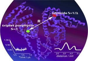
Recently, a new type of spin labels based on photoexcited triplet molecules was proposed for nanometer scale distance measurements by pulsed dipolar electron paramagnetic resonance (PD EPR). However, such molecules are also actively used within biological complexes as photosensitizers for photodynamic therapy (PDT) of cancer. Up to date, the idea of using the photoexcited triplets simultaneously as PDT agents and as spin labels for PD EPR has never been employed. In this work, we demonstrate that PD EPR in conjunction with other methods provides valuable information on the structure and function of PDT candidate complexes, exemplified here with porphyrins bound to human serum albumin (HSA). Two distinct porphyrins with different properties were used: amphiphilic meso-tetrakis(4-hydroxyphenyl)porphyrin (mTHPP) and water soluble cationic meso-tetrakis(N-methyl-4-pyridyl)porphyrin (TMPyP4); HSA was singly nitroxide-labeled to provide a second tag for PD EPR measurements. We found that TMPyP4 locates in a cavity at the center of the four-helix bundle of HSA subdomain IB, close to the interface with solvent, thus being readily accessible to oxygen. As a result, the photolysis of the complex leads to photooxidation of HSA by generated singlet oxygen and causes structural perturbation of the protein. Contrary, in case of mTHPP porphyrin, the binding occurs at the proton-rich pocket of HSA subdomain IIIA, where the access of oxygen to a photosensitizer is hindered. Structural data of PD EPR were supported by other EPR techniques, laser flash photolysis and protein photocleavage studies. Therefore, pulsed EPR on complexes of proteins with photoexcited triplets is a promising approach for gaining structural and functional insights into such PDT agents.
中文翻译:
EPR在卟啉蛋白药物光动力学治疗中的应用。
最近,提出了一种基于光激发三重态分子的新型自旋标记物,用于通过脉冲双极电子顺磁共振(PD EPR)进行纳米尺度距离测量。然而,这些分子也被有效地用于生物复合物中作为光敏剂用于癌症的光动力疗法(PDT)。迄今为止,从未使用将光激发三重态同时用作PDT剂和PD EPR的自旋标记的想法。在这项工作中,我们证明PD EPR与其他方法结合可提供有关PDT候选复合物的结构和功能的有价值信息,此处以结合人血清白蛋白(HSA)的卟啉为例。使用了两种具有不同特性的不同卟啉:两亲性内消旋-四(4-羟苯基)卟啉(mTHPP)和水溶性阳离子内消旋-四(N-甲基-4-吡啶基)卟啉(TMPyP4); HSA单独经过了氮氧化物标记,为PD EPR测量提供了第二个标签。我们发现,TMPyP4位于HSA子域IB的四螺旋束中心的腔中,靠近与溶剂的界面,因此很容易接触到氧气。结果,复合物的光解导致生成的单线态氧对HSA的光氧化,并引起蛋白质的结构扰动。相反,在mTHPP卟啉的情况下,结合发生在HSA亚结构域IIIA的质子丰富的囊袋中,在该处,氧气无法进入光敏剂。PD EPR的结构数据得到其他EPR技术,激光闪光光解和蛋白质光裂研究的支持。因此,


 京公网安备 11010802027423号