当前位置:
X-MOL 学术
›
Stem Cells Transl. Med.
›
论文详情
Our official English website, www.x-mol.net, welcomes your
feedback! (Note: you will need to create a separate account there.)
Magnetic resonance imaging of human neural stem cells in rodent and primate brain.
STEM CELLS Translational Medicine ( IF 5.4 ) Pub Date : 2020-08-25 , DOI: 10.1002/sctm.20-0126 Lisa M McGinley 1 , Matthew S Willsey 2, 3 , Osama N Kashlan 1, 2 , Kevin S Chen 1, 2 , John M Hayes 1 , Ingrid L Bergin 4 , Shayna N Mason 1 , Aaron W Stebbins 1 , Jacquelin F Kwentus 1 , Crystal Pacut 1 , Jennifer Kollmer 5 , Stacey A Sakowski 1 , Caleb B Bell 6, 7 , Cynthia A Chestek 3, 8, 9 , Geoffrey G Murphy 10, 11 , Parag G Patil 1, 2, 3 , Eva L Feldman 1
STEM CELLS Translational Medicine ( IF 5.4 ) Pub Date : 2020-08-25 , DOI: 10.1002/sctm.20-0126 Lisa M McGinley 1 , Matthew S Willsey 2, 3 , Osama N Kashlan 1, 2 , Kevin S Chen 1, 2 , John M Hayes 1 , Ingrid L Bergin 4 , Shayna N Mason 1 , Aaron W Stebbins 1 , Jacquelin F Kwentus 1 , Crystal Pacut 1 , Jennifer Kollmer 5 , Stacey A Sakowski 1 , Caleb B Bell 6, 7 , Cynthia A Chestek 3, 8, 9 , Geoffrey G Murphy 10, 11 , Parag G Patil 1, 2, 3 , Eva L Feldman 1
Affiliation
|
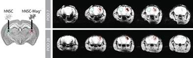
Stem cell transplantation therapies are currently under investigation for central nervous system disorders. Although preclinical models show benefit, clinical translation is somewhat limited by the absence of reliable noninvasive methods to confirm targeting and monitor transplanted cells in vivo. Here, we assess a novel magnetic resonance imaging (MRI) contrast agent derived from magnetotactic bacteria, magneto‐endosymbionts (MEs), as a translatable methodology for in vivo tracking of stem cells after intracranial transplantation. We show that ME labeling provides robust MRI contrast without impairment of cell viability or other important therapeutic features. Labeled cells were visualized immediately post‐transplantation and over time by serial MRI in nonhuman primate and mouse brain. Postmortem tissue analysis confirmed on‐target grft location, and linear correlations were observed between MRI signal, cell engraftment, and tissue ME levels, suggesting that MEs may be useful for determining graft survival or rejection. Overall, these findings indicate that MEs are an effective tool for in vivo tracking and monitoring of cell transplantation therapies with potential relevance to many cellular therapy applications.
中文翻译:

啮齿动物和灵长类动物大脑中人类神经干细胞的磁共振成像。
目前正在研究干细胞移植疗法治疗中枢神经系统疾病。尽管临床前模型显示出益处,但由于缺乏可靠的非侵入性方法来确认靶向和监测体内移植细胞,临床转化在一定程度上受到限制。在这里,我们评估了一种源自趋磁细菌的新型磁共振成像 (MRI) 造影剂,即磁内共生体 (MEs),作为颅内移植后干细胞体内追踪的可翻译方法。我们表明,ME 标记可提供强大的 MRI 对比度,而不会损害细胞活力或其他重要的治疗特征。在移植后立即通过连续 MRI 在非人类灵长类动物和小鼠大脑中观察标记的细胞。尸检组织分析证实了目标移植物位置,在 MRI 信号、细胞移植和组织 ME 水平之间观察到线性相关性,表明 ME 可能有助于确定移植物存活或排斥。总体而言,这些研究结果表明,ME 是一种有效的体内跟踪和监测细胞移植疗法的工具,可能与许多细胞疗法应用相关。
更新日期:2020-08-25
中文翻译:
啮齿动物和灵长类动物大脑中人类神经干细胞的磁共振成像。
目前正在研究干细胞移植疗法治疗中枢神经系统疾病。尽管临床前模型显示出益处,但由于缺乏可靠的非侵入性方法来确认靶向和监测体内移植细胞,临床转化在一定程度上受到限制。在这里,我们评估了一种源自趋磁细菌的新型磁共振成像 (MRI) 造影剂,即磁内共生体 (MEs),作为颅内移植后干细胞体内追踪的可翻译方法。我们表明,ME 标记可提供强大的 MRI 对比度,而不会损害细胞活力或其他重要的治疗特征。在移植后立即通过连续 MRI 在非人类灵长类动物和小鼠大脑中观察标记的细胞。尸检组织分析证实了目标移植物位置,在 MRI 信号、细胞移植和组织 ME 水平之间观察到线性相关性,表明 ME 可能有助于确定移植物存活或排斥。总体而言,这些研究结果表明,ME 是一种有效的体内跟踪和监测细胞移植疗法的工具,可能与许多细胞疗法应用相关。


 京公网安备 11010802027423号