Acta Biomaterialia ( IF 9.4 ) Pub Date : 2020-08-25 , DOI: 10.1016/j.actbio.2020.08.031 Ya Guan 1 , Hong Niu 1 , Yu Dang 1 , Ning Gao 1 , Jianjun Guan 1
|
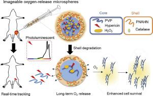
Cell therapy is a promising strategy to treat ischemic diseases, but the efficacy is limited due to high rate of cell death under low oxygen environment of the ischemic tissues. Sustained release of oxygen to continuously oxygenate the transplanted cells may augment cell survival and improve therapeutic efficacy. We have shown previously that oxygen released from oxygen-release microspheres stimulated cell survival in ischemic tissue [1]. To understand how oxygen is released in vivo and duration of release, it is attractive to image the process of oxygen release. Herein, we have developed photoluminenscent oxygen-release microspheres where the in vivo oxygen release can be non-invasively and real-time monitored by an In Vivo Imaging System (IVIS). In the oxygen-release microspheres, a complex of polyvinylpyrrolidone, H2O2 and a fluorescent drug hypericin (HYP) was used as core, and poly(N-isopropylacrylamide-co-acrylate-oligolactide-co-hydroxyethyl methacrylate-co-N-acryloxysuccinimide) conjugated with catalase was used as shell. To distinguish fluorescent signal change for different oxygen release kinetics, the microspheres with various release profiles were developed by using the shell with different degradation rates. In vitro, the fluorescent intensity gradually decreased during the 21-day oxygen release period, consistent with oxygen release kinetics. The released oxygen significantly augmented mesenchymal stem cell (MSC) survival under hypoxic condition. In vivo, the oxygen release rate was faster. The fluorescent signal can be detected for 17 days for the microspheres with the slowest oxygen release kinetics. The implanted microspheres did not induce substantial inflammation. The above results demonstrate that the developed microspheres have potential to monitor the in vivo oxygen release.
中文翻译:
用光致发光的释氧微球对体内的释氧过程进行成像。
细胞疗法是治疗缺血性疾病的一种有前途的策略,但是由于在缺血组织的低氧环境下细胞死亡率高,因此疗效受到限制。持续释放氧气以持续充氧移植的细胞可以延长细胞存活率并提高治疗效果。以前我们已经表明,从氧气释放微球释放的氧气刺激了缺血组织中的细胞存活[1]。为了了解氧气如何在体内释放以及释放的持续时间,对氧气释放过程进行成像很有吸引力。在本文中,我们开发了光致发光的氧释放微球,其中体内的氧释放可以通过体内成像系统(IVIS)进行无创和实时监控。在释氧微球中,聚乙烯吡咯烷酮H 2的配合物Ø 2以荧光药物金丝桃素(HYP)为核,以过氧化氢酶缀合的聚(N-异丙基丙烯酰胺-丙烯酸酯-低聚丙交酯-甲基丙烯酸羟乙酯-甲基丙烯酸-氧代琥珀酰亚胺)-壳为壳。为了区分不同氧气释放动力学的荧光信号变化,使用具有不同降解速率的外壳开发了具有各种释放曲线的微球。在体外,在21天的氧气释放期间,荧光强度逐渐降低,这与氧气释放动力学一致。在低氧条件下,释放的氧气显着增加了间充质干细胞(MSC)的存活率。在体内,氧气释放速度更快。对于氧气释放动力学最慢的微球,可以检测荧光信号17天。植入的微球没有引起实质性炎症。以上结果表明,开发的微球具有监测体内氧气释放的潜力。


 京公网安备 11010802027423号