当前位置:
X-MOL 学术
›
J. Cell. Biochem.
›
论文详情
Our official English website, www.x-mol.net, welcomes your
feedback! (Note: you will need to create a separate account there.)
Exosomes derived from RPE cells under oxidative stress mediate inflammation and apoptosis of normal RPE cells through Apaf1/caspase‐9 axis
Journal of Cellular Biochemistry ( IF 3.0 ) Pub Date : 2020-04-10 , DOI: 10.1002/jcb.29713 Yifeng Ke 1 , Xiaoe Fan 2 , Hao Rui 3 , Ren Xinjun 1 , Wen Dejia 1 , Zheng Chuanzhen 1 , Xiaorong Li 1
Journal of Cellular Biochemistry ( IF 3.0 ) Pub Date : 2020-04-10 , DOI: 10.1002/jcb.29713 Yifeng Ke 1 , Xiaoe Fan 2 , Hao Rui 3 , Ren Xinjun 1 , Wen Dejia 1 , Zheng Chuanzhen 1 , Xiaorong Li 1
Affiliation
|
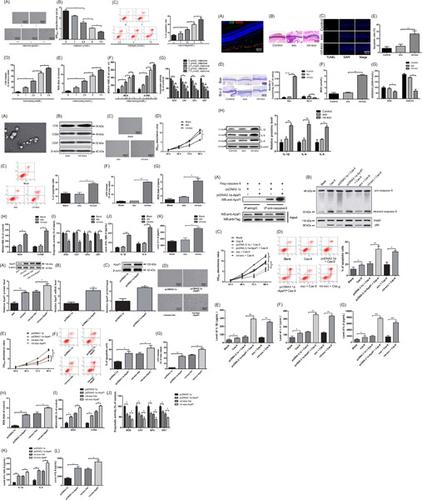
This study aims to explore the effects of exosomes, secreted by retinal pigment epithelial (RPE) cells under oxidative stress (OS), on apoptosis and inflammation of normal RPE cells. Exosomes secreted by normal RPE cells (named as exo) and rotenone (2.5 µmol/L) stimulated RPE cells (named as rot‐exo) were isolated and extracted by multi‐step differential centrifugation for morphology observation under a transmission electron microscopy. pcDNA3.1a, pcDNA3.1a‐Apaf1, and p3xFlag‐CMV‐caspase‐9 plasmids were constructed and transfected into ARPE‐19 cells. Exosomes secreted by ARPE‐19 cells were injected into the vitreous body of rats to verify the effect of Apaf1 and caspase‐9 on cell apoptosis and inflammation. Co‐immunoprecipitation was applied to clarify the interaction of Apaf1 with caspase‐9. Exosomes secreted by rotenone stimulated ARPE‐19 cells could induce cell apoptosis, oxidative injury, and inflammation in ARPE‐19 cells. Exosomes secreted under OS can damage retinal functions of rats and have upregulated expression of Apaf1. Overexpression of Apaf1 in exosomes secreted under OS can cause the inhibition of cell proliferation, the increase of cell apoptosis and elicitation of inflammatory response in ARPE‐19 cells. Exosomes derived from ARPE‐19 cells under OS regulate Apaf1 expression to increase cell apoptosis and to induce oxidative injury and inflammatory response through a caspase‐9 apoptotic pathway.
中文翻译:

氧化应激下RPE细胞衍生的外泌体通过Apaf1/caspase-9轴介导正常RPE细胞的炎症和凋亡
本研究旨在探讨氧化应激(OS)下视网膜色素上皮(RPE)细胞分泌的外泌体对正常RPE细胞凋亡和炎症的影响。通过多步差速离心分离提取正常RPE细胞(命名为exo)和鱼藤酮(2.5 µmol/L)刺激的RPE细胞(命名为rot-exo)分泌的外泌体,并在透射电子显微镜下进行形态观察。构建 pcDNA3.1a、pcDNA3.1a-Apaf1 和 p3xFlag-CMV-caspase-9 质粒并转染到 ARPE-19 细胞中。将 ARPE-19 细胞分泌的外泌体注射到大鼠玻璃体中,验证 Apaf1 和 caspase-9 对细胞凋亡和炎症的影响。应用免疫共沉淀来阐明 Apaf1 与 caspase-9 的相互作用。鱼藤酮刺激 ARPE-19 细胞分泌的外泌体可以诱导 ARPE-19 细胞凋亡、氧化损伤和炎症。OS下分泌的外泌体可以损害大鼠的视网膜功能并上调Apaf1的表达。OS下分泌的外泌体中Apaf1的过度表达可导致ARPE-19细胞增殖抑制、细胞凋亡增加并引发炎症反应。OS 下 ARPE-19 细胞衍生的外泌体调节 Apaf1 表达,增加细胞凋亡,并通过 caspase-9 凋亡途径诱导氧化损伤和炎症反应。
更新日期:2020-04-10
中文翻译:
氧化应激下RPE细胞衍生的外泌体通过Apaf1/caspase-9轴介导正常RPE细胞的炎症和凋亡
本研究旨在探讨氧化应激(OS)下视网膜色素上皮(RPE)细胞分泌的外泌体对正常RPE细胞凋亡和炎症的影响。通过多步差速离心分离提取正常RPE细胞(命名为exo)和鱼藤酮(2.5 µmol/L)刺激的RPE细胞(命名为rot-exo)分泌的外泌体,并在透射电子显微镜下进行形态观察。构建 pcDNA3.1a、pcDNA3.1a-Apaf1 和 p3xFlag-CMV-caspase-9 质粒并转染到 ARPE-19 细胞中。将 ARPE-19 细胞分泌的外泌体注射到大鼠玻璃体中,验证 Apaf1 和 caspase-9 对细胞凋亡和炎症的影响。应用免疫共沉淀来阐明 Apaf1 与 caspase-9 的相互作用。鱼藤酮刺激 ARPE-19 细胞分泌的外泌体可以诱导 ARPE-19 细胞凋亡、氧化损伤和炎症。OS下分泌的外泌体可以损害大鼠的视网膜功能并上调Apaf1的表达。OS下分泌的外泌体中Apaf1的过度表达可导致ARPE-19细胞增殖抑制、细胞凋亡增加并引发炎症反应。OS 下 ARPE-19 细胞衍生的外泌体调节 Apaf1 表达,增加细胞凋亡,并通过 caspase-9 凋亡途径诱导氧化损伤和炎症反应。


 京公网安备 11010802027423号