当前位置:
X-MOL 学术
›
J. Synchrotron Radiat.
›
论文详情
Our official English website, www.x-mol.net, welcomes your feedback! (Note: you will need to create a separate account there.)
Multiscale pink-beam microCT imaging at the ESRF-ID17 biomedical beamline.
Journal of Synchrotron Radiation ( IF 2.5 ) Pub Date : 2020-08-18 , DOI: 10.1107/s160057752000911x Alberto Mittone 1 , Luca Fardin 2 , Francesca Di Lillo 2 , Michela Fratini 3 , Herwig Requardt 2 , Anthony Mauro 2 , Roberto Arturo Homs-Regojo 2 , Paul Antoine Douissard 2 , Giacomo E Barbone 4 , Johannes Stroebel 4 , Mariele Romano 4 , Lorenzo Massimi 3 , Ginevra Begani-Provinciali 3 , Francesca Palermo 3 , Sam Bayat 5 , Alessia Cedola 3 , Paola Coan 4 , Alberto Bravin 2
Journal of Synchrotron Radiation ( IF 2.5 ) Pub Date : 2020-08-18 , DOI: 10.1107/s160057752000911x Alberto Mittone 1 , Luca Fardin 2 , Francesca Di Lillo 2 , Michela Fratini 3 , Herwig Requardt 2 , Anthony Mauro 2 , Roberto Arturo Homs-Regojo 2 , Paul Antoine Douissard 2 , Giacomo E Barbone 4 , Johannes Stroebel 4 , Mariele Romano 4 , Lorenzo Massimi 3 , Ginevra Begani-Provinciali 3 , Francesca Palermo 3 , Sam Bayat 5 , Alessia Cedola 3 , Paola Coan 4 , Alberto Bravin 2
Affiliation
|
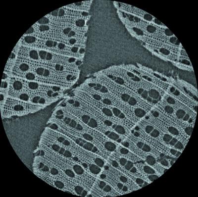
Recent trends in hard X‐ray micro‐computed tomography (microCT) aim at increasing both spatial and temporal resolutions. These challenges require intense photon beams. Filtered synchrotron radiation beams, also referred to as `pink beams', which are emitted by wigglers or bending magnets, meet this need, owing to their broad energy range. In this work, the new microCT station installed at the biomedical beamline ID17 of the European Synchrotron is described and an overview of the preliminary results obtained for different biomedical‐imaging applications is given. This new instrument expands the capabilities of the beamline towards sub‐micrometre voxel size scale and simultaneous multi‐resolution imaging. The current setup allows the acquisition of tomographic datasets more than one order of magnitude faster than with a monochromatic beam configuration.
中文翻译:

ESRF-ID17生物医学光束线的多尺度粉红光束microCT成像。
硬X射线微计算机断层扫描(microCT)的最新趋势旨在提高空间和时间分辨率。这些挑战需要强烈的光子束。摆动的或弯曲的磁体发出的经滤波的同步加速器辐射束(也称为“粉红色束”)由于其广泛的能量范围而满足了这一需求。在这项工作中,描述了安装在欧洲同步加速器生物医学射线线ID17上的新microCT站,并概述了针对不同生物医学成像应用获得的初步结果。这款新仪器将光束线的功能扩展到亚微米体素尺寸刻度和同步多分辨率成像。
更新日期:2020-08-18
中文翻译:
ESRF-ID17生物医学光束线的多尺度粉红光束microCT成像。
硬X射线微计算机断层扫描(microCT)的最新趋势旨在提高空间和时间分辨率。这些挑战需要强烈的光子束。摆动的或弯曲的磁体发出的经滤波的同步加速器辐射束(也称为“粉红色束”)由于其广泛的能量范围而满足了这一需求。在这项工作中,描述了安装在欧洲同步加速器生物医学射线线ID17上的新microCT站,并概述了针对不同生物医学成像应用获得的初步结果。这款新仪器将光束线的功能扩展到亚微米体素尺寸刻度和同步多分辨率成像。


 京公网安备 11010802027423号