Structure ( IF 4.4 ) Pub Date : 2020-08-18 , DOI: 10.1016/j.str.2020.07.017 Gong-Her Wu 1 , Patrick G Mitchell 2 , Jesus G Galaz-Montoya 1 , Corey W Hecksel 2 , Emily M Sontag 3 , Vimal Gangadharan 4 , Jeffrey Marshman 4 , David Mankus 5 , Margaret E Bisher 5 , Abigail K R Lytton-Jean 5 , Judith Frydman 3 , Kirk Czymmek 6 , Wah Chiu 7
|
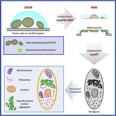
Three-dimensional (3D) visualization of vitrified cells can uncover structures of subcellular complexes without chemical fixation or staining. Here, we present a pipeline integrating three imaging modalities to visualize the same specimen at cryogenic temperature at different scales: cryo-fluorescence confocal microscopy, volume cryo-focused ion beam scanning electron microscopy, and transmission cryo-electron tomography. Our proof-of-concept benchmark revealed the 3D distribution of organelles and subcellular structures in whole heat-shocked yeast cells, including the ultrastructure of protein inclusions that recruit fluorescently-labeled chaperone Hsp104. Since our workflow efficiently integrates imaging at three different scales and can be applied to other types of cells, it could be used for large-scale phenotypic studies of frozen-hydrated specimens in a variety of healthy and diseased conditions with and without treatments.
中文翻译:
玻璃化细胞的多尺度 3D 冷冻相关显微镜。
玻璃化细胞的三维 (3D) 可视化可以揭示亚细胞复合物的结构,无需化学固定或染色。在这里,我们提出了一个集成三种成像模式的管道,以在不同尺度的低温下可视化同一样本:低温荧光共焦显微镜、体积低温聚焦离子束扫描电子显微镜和透射低温电子断层扫描。我们的概念验证基准揭示了整个热休克酵母细胞中细胞器和亚细胞结构的 3D 分布,包括招募荧光标记伴侣 Hsp104 的蛋白质内含物的超微结构。由于我们的工作流程有效地集成了三种不同尺度的成像,并且可以应用于其他类型的细胞,因此它可以用于在各种健康和患病条件下进行或未经治疗的冷冻水合样本的大规模表型研究。


 京公网安备 11010802027423号