Biochimica et Biophysica Acta (BBA) - General Subjects ( IF 2.8 ) Pub Date : 2020-08-15 , DOI: 10.1016/j.bbagen.2020.129708 Pornpattra Maphanao 1 , Raynoo Thanan 1 , Watcharin Loilome 1 , Sirinart Chio-Srichan 2 , Molin Wongwattanakul 3 , Chadamas Sakonsinsiri 1
|
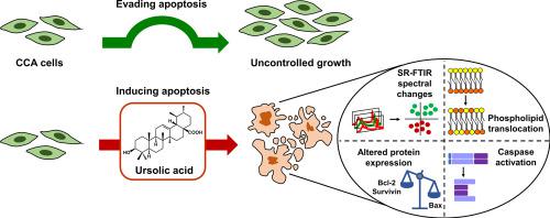
Background
Ursolic acid (UA) is a natural triterpenoid which possesses anti-cancer activity. However, little is known regarding the activity and molecular mechanism of UA in cholangiocarcinoma (CCA). Thus, we investigated the effects of UA on growth inhibition and apoptosis induction through biomolecular changes in KKU-213 and KKU-055 CCA cell lines.
Methods
The anti-proliferative effect of UA against CCA cells was evaluated using SRB assay. Changes in biomolecules were assessed by SR-FTIR microspectroscopy combined with PCA and conventional methods (i.e., Annexin V-FITC/PI staining for lipid alteration and apoptosis induction; Western blot analysis and caspase-3/7 activity assay for apoptotic protein detection).
Results
UA suppressed the proliferation of CCA cells in a dose- and time-dependent manner. SR-FTIR data revealed a significant alteration in lipids attributable to changes in apoptotic cell membranes, confirmed by Annexin V-FITC/PI staining. SR-FTIR data showed that UA promoted changes in the protein secondary structure. Elevated expression of Bax and decreased expression of Bcl-2 and survivin/BIRC5 along with augmented caspase-3/7 activity supported alterations in apoptosis-related proteins.
Conclusions
SR-FTIR microspectroscopy was successfully used as a label-free technique to monitor apoptosis-induced biomolecular changes in UA-treated CCA cells. UA exerted the cytotoxic and apoptotic activities in CCA cells through alterations in membrane lipids and apoptotic proteins. UA could be a potential anti-CCA candidate and a chemical starting point for the discovery of novel anti-cancer agents.
Significance
Our present study showed the first evidence that UA exhibited the anti-proliferative and pro-apoptotic activities toward CCA cells through changes in biomolecules, notably lipids and proteins.
中文翻译:
同步加速器 FTIR 显微光谱揭示了熊果酸处理后的胆管癌细胞凋亡诱导的生物分子变化。
背景
熊果酸(UA)是一种天然三萜类化合物,具有抗癌活性。然而,关于 UA 在胆管癌 (CCA) 中的活性和分子机制知之甚少。因此,我们通过 KKU-213 和 KKU-055 CCA 细胞系的生物分子变化研究了 UA 对生长抑制和凋亡诱导的影响。
方法
使用SRB测定评估UA对CCA细胞的抗增殖作用。通过SR-FTIR显微光谱结合PCA和常规方法(即Annexin V-FITC/PI染色用于脂质改变和凋亡诱导;Western blot分析和caspase-3/7活性测定用于凋亡蛋白检测)评估生物分子的变化。
结果
UA 以剂量和时间依赖性方式抑制 CCA 细胞的增殖。 SR-FTIR 数据显示,由于凋亡细胞膜的变化,脂质发生了显着变化,这经膜联蛋白 V-FITC/PI 染色证实。 SR-FTIR数据表明UA促进了蛋白质二级结构的变化。 Bax 表达升高、Bcl-2 和 survivin/BIRC5 表达降低以及 caspase-3/7 活性增强支持细胞凋亡相关蛋白的改变。
结论
SR-FTIR 显微光谱学已成功用作无标记技术来监测 UA 处理的 CCA 细胞中凋亡诱导的生物分子变化。 UA 通过改变膜脂和凋亡蛋白在 CCA 细胞中发挥细胞毒性和凋亡活性。 UA 可能是一种潜在的抗 CCA 候选药物,也是发现新型抗癌药物的化学起点。
意义
我们目前的研究首次证明 UA 通过改变生物分子,特别是脂质和蛋白质,对 CCA 细胞表现出抗增殖和促凋亡活性。


 京公网安备 11010802027423号