Our official English website, www.x-mol.net, welcomes your
feedback! (Note: you will need to create a separate account there.)
Quantitative analysis of fluorescent ligand binding to dopamine D3 receptors using live‐cell microscopy
The FEBS Journal ( IF 5.5 ) Pub Date : 2020-08-11 , DOI: 10.1111/febs.15519 Anni Allikalt 1, 2 , Tõnis Laasfeld 1 , Mihkel Ilisson 1 , Sergei Kopanchuk 1 , Ago Rinken 1
The FEBS Journal ( IF 5.5 ) Pub Date : 2020-08-11 , DOI: 10.1111/febs.15519 Anni Allikalt 1, 2 , Tõnis Laasfeld 1 , Mihkel Ilisson 1 , Sergei Kopanchuk 1 , Ago Rinken 1
Affiliation
|
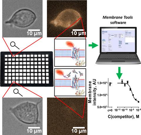
Dopamine receptors are G protein‐coupled receptors that have several essential functions in the central nervous system. A better understanding of the regulatory mechanisms of ligand binding to the receptor may open new possibilities to affect the downstream signal transduction pathways. The majority of the available ligand binding assays use either membrane preparations, cell suspensions, or genetically modified receptors, which may give at least partially incorrect understanding of ligand binding. In this study, we implemented an assay combining fluorescence and bright‐field microscopy to measure ligand binding to dopamine D3 receptors in live mammalian cells. For membrane fluorescence intensity quantification from microscopy images, we developed a machine learning‐based user‐friendly software membrane tools and incorporated it into a data management software aparecium that has been previously developed in our workgroup. For the experiments, a fluorescent ligand NAPS‐Cy3B was synthesized by conjugating a dopaminergic antagonist N‐(p‐aminophenethyl)spiperone with a fluorophore Cy3B. The subnanomolar affinity of NAPS‐Cy3B makes it a suitable ligand for the characterization of D3 receptors in live HEK293 cells. Using a microplate compatible automated widefield fluorescence microscope, together with the membrane tools software, enables the detection and quantification of ligand binding with a high‐throughput. The live cell assay is suitable for the characterization of fluorescent ligand binding and also in the competition experiments for the screening of novel unlabeled dopaminergic ligands. We propose that this simple yet more native‐like approach is feasible in GPCR research, as it enables the detection of ligand binding in an environment containing more components involved in the signal transduction cascade.
中文翻译:

使用活细胞显微镜对荧光配体与多巴胺D3受体结合的定量分析
多巴胺受体是G蛋白偶联的受体,在中枢神经系统中具有多种基本功能。对配体与受体结合的调节机制的更好理解可能会为影响下游信号转导途径开辟新的可能性。大多数可用的配体结合测定使用膜制剂,细胞悬液或基因修饰的受体,这可能至少部分使配体结合理解不正确。在这项研究中,我们实施了一种结合荧光和明视野显微镜的检测方法,以测量配体与活哺乳动物细胞中多巴胺D 3受体的结合。为了从显微镜图像定量分析膜荧光强度,我们开发了一种基于机器学习的用户友好型软件膜工具并将其整合到先前在我们的工作组中开发的数据管理软件aparecium中。在实验中,通过将多巴胺能拮抗剂N-(对氨基苯乙基)哌酮与荧光团Cy3B缀合,合成了荧光配体NAPS-Cy3B。NAPS-Cy3B的亚纳摩尔亲和力使其成为表征活HEK293细胞中D 3受体的合适配体。使用与微板兼容的自动宽视野荧光显微镜以及膜工具该软件可实现高通量检测和定量配体结合。活细胞测定适用于荧光配体结合的表征,也适用于筛选新型未标记多巴胺能配体的竞争实验。我们建议,这种简单但更像本机的方法在GPCR研究中是可行的,因为它可以在包含更多涉及信号转导级联的组分的环境中检测配体结合。
更新日期:2020-08-11
中文翻译:
使用活细胞显微镜对荧光配体与多巴胺D3受体结合的定量分析
多巴胺受体是G蛋白偶联的受体,在中枢神经系统中具有多种基本功能。对配体与受体结合的调节机制的更好理解可能会为影响下游信号转导途径开辟新的可能性。大多数可用的配体结合测定使用膜制剂,细胞悬液或基因修饰的受体,这可能至少部分使配体结合理解不正确。在这项研究中,我们实施了一种结合荧光和明视野显微镜的检测方法,以测量配体与活哺乳动物细胞中多巴胺D 3受体的结合。为了从显微镜图像定量分析膜荧光强度,我们开发了一种基于机器学习的用户友好型软件膜工具并将其整合到先前在我们的工作组中开发的数据管理软件aparecium中。在实验中,通过将多巴胺能拮抗剂N-(对氨基苯乙基)哌酮与荧光团Cy3B缀合,合成了荧光配体NAPS-Cy3B。NAPS-Cy3B的亚纳摩尔亲和力使其成为表征活HEK293细胞中D 3受体的合适配体。使用与微板兼容的自动宽视野荧光显微镜以及膜工具该软件可实现高通量检测和定量配体结合。活细胞测定适用于荧光配体结合的表征,也适用于筛选新型未标记多巴胺能配体的竞争实验。我们建议,这种简单但更像本机的方法在GPCR研究中是可行的,因为它可以在包含更多涉及信号转导级联的组分的环境中检测配体结合。


 京公网安备 11010802027423号