当前位置:
X-MOL 学术
›
NMR Biomed.
›
论文详情
Our official English website, www.x-mol.net, welcomes your
feedback! (Note: you will need to create a separate account there.)
A pilot study of magnetic resonance fingerprinting in Parkinson's disease.
NMR in Biomedicine ( IF 2.7 ) Pub Date : 2020-08-11 , DOI: 10.1002/nbm.4389 Vera Catharina Keil 1, 2 , Stilyana Peteva Bakoeva 1, 3 , Alina Jurcoane 1, 4 , Mariya Doneva 5 , Thomas Amthor 5 , Peter Koken 5 , Burkhard Mädler 6 , Guido Lüchters 7 , Wolfgang Block 8 , Ullrich Wüllner 9, 10 , Elke Hattingen 1, 4
NMR in Biomedicine ( IF 2.7 ) Pub Date : 2020-08-11 , DOI: 10.1002/nbm.4389 Vera Catharina Keil 1, 2 , Stilyana Peteva Bakoeva 1, 3 , Alina Jurcoane 1, 4 , Mariya Doneva 5 , Thomas Amthor 5 , Peter Koken 5 , Burkhard Mädler 6 , Guido Lüchters 7 , Wolfgang Block 8 , Ullrich Wüllner 9, 10 , Elke Hattingen 1, 4
Affiliation
|
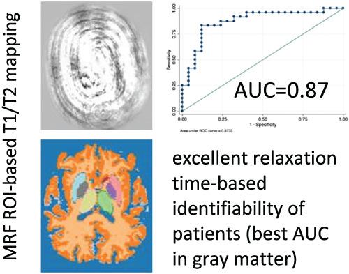
Parkinson's disease (PD) affects more than six million people, but reliable MRI biomarkers with which to diagnose patients have not been established. Magnetic resonance fingerprinting (MRF) is a recent quantitative technique that can provide relaxometric maps from a single sequence. The purpose of this study is to assess the potential of MRF to identify PD in patients and their disease severity, as well as to evaluate comfort during MRF. Twenty‐five PD patients and 25 matching controls underwent 3 T MRI, including an axial 2D spoiled gradient echo MRF sequence. T1 and T2 maps were generated by voxel‐wise matching the measured MRF signal to a precomputed dictionary. All participants also received standard inversion recovery T1 and multi‐echo T2 mapping. An ROI‐based analysis of relaxation times was performed. Differences between patients and controls as well as techniques were determined by logistic regression, Spearman correlation and t‐test. Patients were asked to estimate the subjective comfort of the MRF sequence. Both MRF‐based T1 and T2 mapping discriminated patients from controls: T1 relaxation times differed most in cortical grey matter (PD 1337 ± 38 vs. control 1386 ± 37 ms; mean ± SD; P = .0001) and, in combination with normal‐appearing white matter, enabled correct discrimination in 85.7% of cases (sensitivity 83.3%; specificity 88.0%; receiver‐operating characteristic [ROC]) area under the curve [AUC] 0.87), while for T2 mapping the left putamen was the strongest classifier (40.54 ± 6.28 vs. 34.17 ± 4.96 ms; P = .0001), enabling differentiation of groups in 84.0% of all cases (sensitivity 80.0%; specificity 88.0%; ROC AUC 0.87). Relaxation time differences were not associated with disease severity. Standard mapping techniques generated significantly different relaxation time values and identified other structures as different between groups other than MRF. Twenty‐three out of 25 PD patients preferred the MRF examination instead of a standard MRI. MRF‐based mapping can identify PD patients with good comfort but needs further assessment regarding disease severity identification and its potential for comparability with standard mapping technique results.
中文翻译:

帕金森病磁共振指纹图谱的初步研究。
帕金森病 (PD) 影响超过 600 万人,但尚未建立可靠的 MRI 生物标志物来诊断患者。磁共振指纹图谱 (MRF) 是一种最新的定量技术,可以从单个序列提供弛豫图。本研究的目的是评估 MRF 在识别患者 PD 及其疾病严重程度方面的潜力,以及评估 MRF 期间的舒适度。25 名 PD 患者和 25 名匹配对照接受了 3 T MRI,包括轴向 2D 破坏梯度回波 MRF 序列。T1 和 T2 图是通过将测量的 MRF 信号与预先计算的字典进行体素匹配来生成的。所有参与者还接受了标准倒置恢复 T1 和多回波 T2 映射。进行了基于 ROI 的弛豫时间分析。通过逻辑回归、Spearman 相关性和 t 检验确定患者和对照以及技术之间的差异。要求患者估计 MRF 序列的主观舒适度。基于 MRF 的 T1 和 T2 映射都将患者与对照组区分开来:T1 松弛时间在皮质灰质中差异最大(PD 1337 ± 38 与对照 1386 ± 37 ms;平均值 ± SD;P = .0001) 并且与外观正常的白质相结合,能够正确区分 85.7% 的病例(敏感性 83.3%;特异性 88.0%;受试者工作特征 [ROC])曲线下面积 [AUC] 0.87) ,而对于 T2 映射,左壳核是最强的分类器(40.54 ± 6.28 对 34.17 ± 4.96 ms;P= .0001),使所有病例的 84.0% 能够区分组(敏感性 80.0%;特异性 88.0%;ROC AUC 0.87)。松弛时间差异与疾病严重程度无关。标准映射技术产生了显着不同的弛豫时间值,并确定了除 MRF 之外的组之间不同的其他结构。25 名 PD 患者中有 23 名更喜欢 MRF 检查而不是标准 MRI。基于 MRF 的映射可以识别具有良好舒适度的 PD 患者,但需要进一步评估疾病严重程度识别及其与标准映射技术结果的可比性。
更新日期:2020-10-05
中文翻译:
帕金森病磁共振指纹图谱的初步研究。
帕金森病 (PD) 影响超过 600 万人,但尚未建立可靠的 MRI 生物标志物来诊断患者。磁共振指纹图谱 (MRF) 是一种最新的定量技术,可以从单个序列提供弛豫图。本研究的目的是评估 MRF 在识别患者 PD 及其疾病严重程度方面的潜力,以及评估 MRF 期间的舒适度。25 名 PD 患者和 25 名匹配对照接受了 3 T MRI,包括轴向 2D 破坏梯度回波 MRF 序列。T1 和 T2 图是通过将测量的 MRF 信号与预先计算的字典进行体素匹配来生成的。所有参与者还接受了标准倒置恢复 T1 和多回波 T2 映射。进行了基于 ROI 的弛豫时间分析。通过逻辑回归、Spearman 相关性和 t 检验确定患者和对照以及技术之间的差异。要求患者估计 MRF 序列的主观舒适度。基于 MRF 的 T1 和 T2 映射都将患者与对照组区分开来:T1 松弛时间在皮质灰质中差异最大(PD 1337 ± 38 与对照 1386 ± 37 ms;平均值 ± SD;P = .0001) 并且与外观正常的白质相结合,能够正确区分 85.7% 的病例(敏感性 83.3%;特异性 88.0%;受试者工作特征 [ROC])曲线下面积 [AUC] 0.87) ,而对于 T2 映射,左壳核是最强的分类器(40.54 ± 6.28 对 34.17 ± 4.96 ms;P= .0001),使所有病例的 84.0% 能够区分组(敏感性 80.0%;特异性 88.0%;ROC AUC 0.87)。松弛时间差异与疾病严重程度无关。标准映射技术产生了显着不同的弛豫时间值,并确定了除 MRF 之外的组之间不同的其他结构。25 名 PD 患者中有 23 名更喜欢 MRF 检查而不是标准 MRI。基于 MRF 的映射可以识别具有良好舒适度的 PD 患者,但需要进一步评估疾病严重程度识别及其与标准映射技术结果的可比性。


 京公网安备 11010802027423号